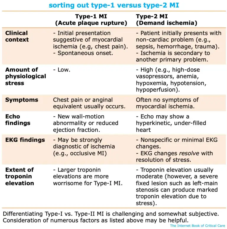
Subjective: Patient usually presents with noncardiac problems (e.g., pneumonia, sepsis, DKA, gastroenteritis).
ECG: Usually will show features of NOMI.
Exam: POCUS may be reassuring (e.g., hyperkinetic heart without wall-motion abnormality).
Other tests: Troponin elevation (but usually less than with type-I MI).
PE causing pleural infarct with chest pain
Subjective: Sharp, pleuritic chest pain.
ECG: Often unchanged.
Exam: POCUS may show pleural effusion, small area of lung consolidation, and possibly DVT.
Other tests: D-dimer elevation; focal opacity may be seen on chest X-ray; CT angiography is diagnostic.
PE causing RV strain with myocardial ischemia
Subjective: May cause ischemic-quality anginal chest pain due to hypoperfusion of the right ventricular free wall. Patients often present with (pre)syncope or dyspnea.
ECG may show:
T-wave inversion involving the interior leads and/or right precordial leads.
Right axis deviation (RAD).
Complete or partial right bundle branch block (RBBB).
SI-QIII-TIII pattern.
ST elevation rarely seen (aVR).
Tachycardia.
Exam: POCUS should show RV dilation and RV systolic dysfunction. DVT study may be positive.
Other tests: CT angiography should be diagnostic.
💡 (Sub)massive PE causing ischemia of the right ventricular free wall can closely mimic MI (e.g., with anginal-type chest pain, positive troponin, and dynamic ECG changes).
aortic dissection
Subjective: Chest pain (sharp, tearing/ripping, rapid acceleration to maximal pain, radiation to shoulders, migratory). May be associated with a variety of symptoms beyond the chest (e.g., neurologic, abdominal pain).
ECG: Usually nonspecific. Aortic dissection may rarely cause occlusion of a coronary artery, producing an OMI-pattern ECG.
Exam: Findings vary based on location of dissection, potentially including:
Bp differential between limbs.
POCUS: Aortic root dilation, aortic regurgitation, pericardial effusion. Ultrasound of abdominal aorta, carotids, or femoral arteries may show a dissection flap.
Other tests: Chest X-ray may show widening of mediastinum. Dissection protocol CT angiography should be diagnostic.
pericarditis
Subjective: Chest pain is often positional (worse lying flat), pleuritic, and may be sharp. Pain may be associated with fever and flu-like symptoms.
ECG: May see diffuse ST elevation or T-wave changes.
Exam: Pericardial friction rub may be heard. Echo may show pericardial effusion.
pneumothorax
Subjective: Sharp pleuritic pain, dyspnea.
Exam: Subcutaneous emphysema may occur. Lung ultrasonography should be diagnostic.
Other tests: Chest X-ray or chest CT scan in more complex cases.