Pearls
- lack of objective gait abnormality has 99% NPV for dizzy stroke
Strategy:
In the ED
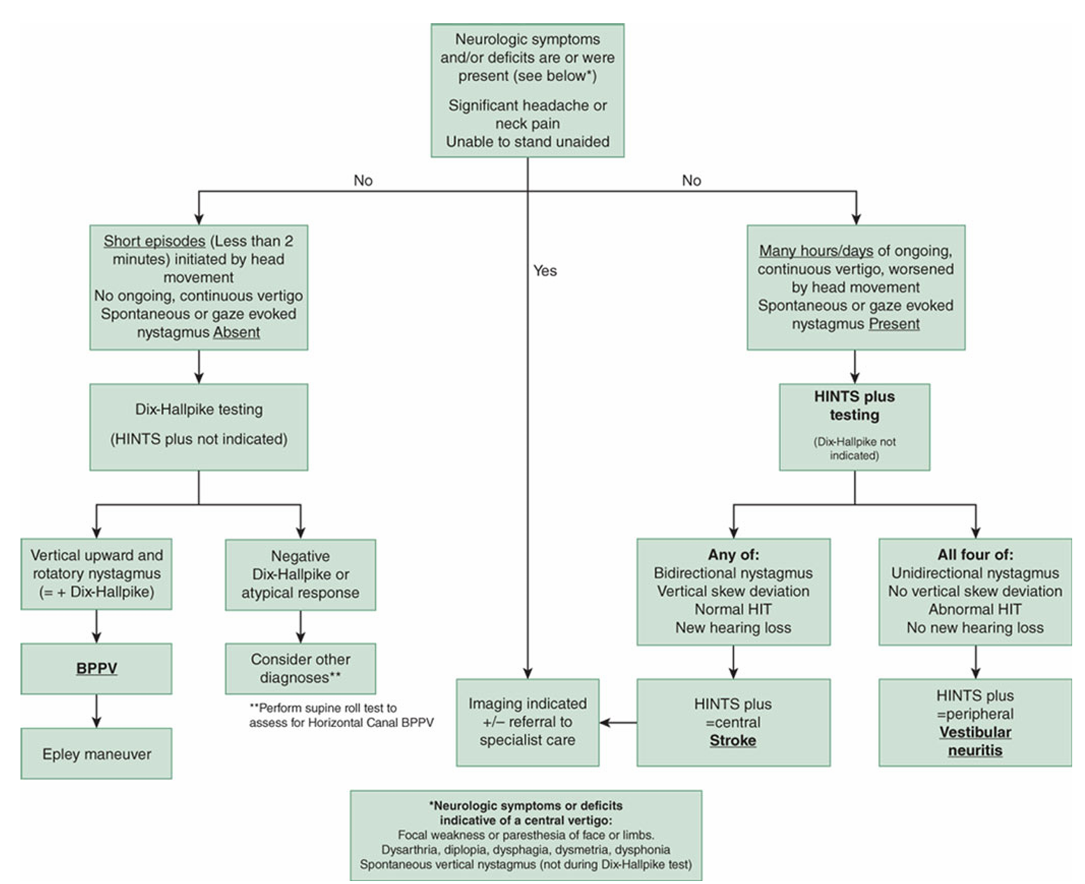
Continuous Vertigo + Nystagmus at Rest?
- Rule out central causes
- New headache or neck pain?
- Dangerous D’s:
- EOM’s
- Speech:
- Language Comprehension
- Perform gait assessment
- New objective gait abnormality or trunkal ataxia?
- Perform HINTS
Acute Vestibular Syndrome
DDx:
- Vestibular Neuritis
- Posterior Stroke
- BPPV
- Vestibular Migraine
Signs/symptoms:
- Vertigo
- Nystagmus
- Nausea/vomiting
- Head-motion intolerance
- unsteady gait
BPPV
- <2 min episode of dizziness
- brought on by position change
- no nystagmus or dizziness at rest
- Dix Hallpike Test ->
- Latency (nothing happens for a few seconds)
- crescendo->decrescendo vertical upward/torsional nystagmus (10-30s)
- purely horizontal nystagmus -> think HC BPPV
- Affected ear is downward ear, other side is normal
Vestibular Migraine
- Multiple dizzy spells lasting mins/hours/days
- Hx of migraine headaches
- Most spells have migraine features
- Dix Hallpike Test-> none or not typical
CANNOT see:
- bidirectional nystagmus (changes beating direction with gaze direction)
- vertical or diagonal skew
MUST see: - nystagmus that doesn’t change direction with gaze
- catch up saccade
HINTS
- MUST have constant dizziness and nystagmus at rest with NO central features
- HINTS plus
- all of the above PLUS no new hearing loss (worrisome for AICA stroke)
Diagnostic Approach to Dizzy Patients
- Screen for central features
- new headache or neck pain
- focal weakness or paresthesia
- Diplopia, dysarthria, dysmetria, dysphonia, dysphagia
- Vertical nystagmus at rest
- New inability to walk unaided (objective gait abnormality)
- Evaluate patient history for BPPV and Vestibular Neuritis
- BPPV: Episodic, triggered by position change, no nystagmus at rest
- Perform Dix Hallpike Test
- Vestibular Neuritis: Constant dizziness (hours/days), worsened by position change, nystagmus at rest
- Perform HINTS exam
- Patient fixates gaze on a target
- Loose back and forth, the rapid back to center (at least 20 degrees )
- fast component of nystagmus beats away from affected ear
- abnormal HIT seen when head is turned towards affected ear (catch up saccade)
- RIGHT beating nystagmus will have an abnormal LEFT HIT
- Perform HINTS exam
- BPPV: Episodic, triggered by position change, no nystagmus at rest
- Ruling IN BPPV / VN = Ruling OUT stroke
- If positive for central features or HINTS central
- work up for stroke
Treatment
Research
- Objective Gait Abnormality: 99% NPV for Posterior Stroke
- Clinical diagnosis of benign paroxysmal positional vertigo and vestibular neuritis | CMAJ
- GRACE-3 Dizziness - Emergency Medicine Clinical Practice Guideline | SAEM
Vertigo/Dizziness Types and DDx
- Acute Prolonged
- Vestibular Neuritis
- Stroke
- Recurrent Spontaneous
- Meniere’s
- Vestibular Migraine
- Psychogenic Dizziness
- Vertebrobasilar TIA
- Recurrent Positional
- BPPV
- Chronic Persistent
- Neurodegenerative Disorders
Vestibular Neuritis
- Nystagmus beating one direction
- no vertical skew
- Catch up saccade when head turned rapidly
- No new hearing loss
Central Features
- new headache or neck pain
- focal weakness or paresthesia
- Diplopia, dysarthria, dysmetria, dysphonia, dysphagia,
- Vertical nystagmus at rest
- New ability to walk unaided
As I state in the video, first I screen for central features.
Those that screen positive get a work up.
If they screen negative, and have no nystagmus and sounds like it could be BPPV, do the Dix-Hallpike test. If characteristic nystagmus of posterior canal BPPV is seen, do an Epley.
Then if they have nystagmus, I do the HINTS exam. (and check for hearing loss)
Those with HINTS central result, get a work up.
Those without nystagmus I walk. If they have a new objective difficulty walking, they get a work up.
Those without an new objective difficulty walking, consider general medical causes of dizziness. (hypoNa, UTI, CO poisoning etc.)
Patients who are older can usually participate in the exam. Patients who are confused (if new confusion, work them up). If they are already confused, well, they are difficult to evaluate for any complaint. Do the best you can.
When I was in practice, I rarely ordered MRI’s.
Most posterior circulations strokes have central features. Most patients with nystagmus that don’t, have vestibular neuritis and can be sent home if they have a HINTS peripheral exam.
Physiology
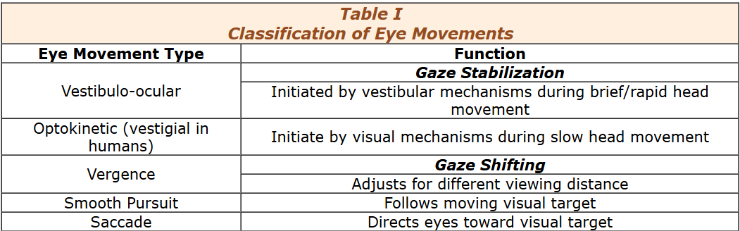
- Vestibular nystagmus is a vestibulo-ocular response - driven by a vestibular stimulus (i.e., accelerating head movement or percieved movememnt)
Neuro-ophthalmology / Physiology of Conjugate Gaze Deviation in Stroke
- Damage to the frontal cortical eye field and the midbrain (superior colliculus) effect voluntary and reflex saccades, particularly those in the horizontal plane.
- Immediately following unilateral damage of the frontal cortical eye field, there is an inability to voluntarily initiate a horizontal eye movement in a direction contralateral to (away from) the side of the lesion.
- i.e. immediately following a right frontal lobe lesion, both eyes cannot be moved voluntarily to the left beyond the midline
- However, both eyes will move to the left beyond the midline to vestibular stimulation.
- The deficits disappear with time if the damage is localized to the frontal cortical eye field and does not involve the superior colliculus.
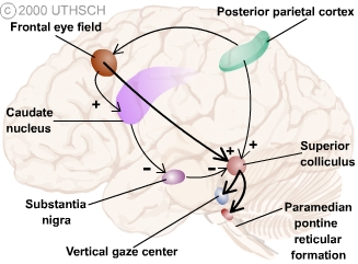
Frontal Eye Fields
- are involved in initiating voluntary saccades that locate and focus on a particular object-of-interest.
- are located posteriorly in the middle frontal gyrus.
- send their axons in the internal capsule, crus cerebri and corticotectal tract to the midbrain where they decussate and end in the superior colliculus.
Horizontal Gaze Center
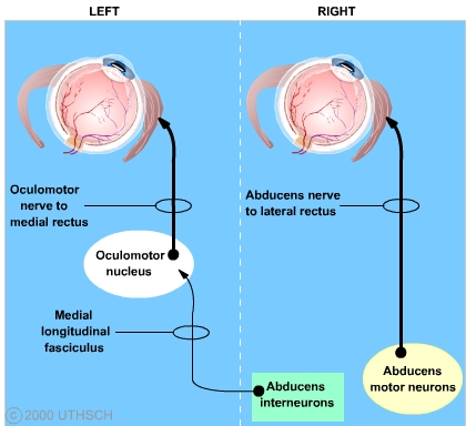
- is called the paramedian pontine reticular formation (PPRF)
- has direct control over the abducens lower motor neurons and interneurons
- Recall that the abducens nucleus contains
- lower motor neurons that send their axons in the ipsilateral abducens nerve to the lateral rectus muscle
- interneurons that send their axons in the contralateral medial longitudinal fasciculus to the oculomotor neurons controlling the medial rectus
- Recall that the abducens nucleus contains
Superior Colliculus
Can initiate and control saccades independent of input from the frontal eye field. However, the motor control signals initiated by the frontal eye field and the superior colliculus differ in function.
- Normally the frontal eye field initiates voluntary and memory-guided saccades,
- the superior colliculus initiates reflex orienting saccades
Test
I. Core Principle
Patients who screen negative for central features, have no objective gait deficit, and have peripheral-pattern findings (BPPV or vestibular neuritis) are extremely unlikely to have a cerebellar/brainstem stroke.
II. Categorizing Dizziness
Use a timing & triggers framework:
| Syndrome | Typical Etiology |
|---|---|
| Acute Prolonged (continuous hours–days) | Vestibular neuritis, posterior stroke |
| Recurrent Positional (brief, triggered) | BPPV |
| Recurrent Spontaneous | Vestibular migraine, Meniere’s, psychogenic, VBTIA |
| Chronic Persistent | Neurodegenerative, chronic vestibulopathy |
III. ED Algorithm
Step 1 — Screen for Central Features (STOP if present)
Any of these → do not perform HINTS, move directly to stroke workup.
Central red flags
-
New headache or neck pain
-
Focal neurologic deficits: weakness, sensory loss
-
Cranial nerve or cerebellar signs:
- Diplopia
- Dysarthria
- Dysmetria
- Dysphonia
- Dysphagia
-
Vertical nystagmus at rest
-
Bidirectional gaze-evoked nystagmus
-
New inability to walk unaided (objective gait ataxia)
If any central feature is present → consider posterior circulation stroke until proven otherwise.
Step 2 — Is the dizziness continuous with nystagmus at rest?
If YES → Acute Vestibular Syndrome (AVS)
Go to HINTS pathway.
If NO, then evaluate for episodic triggers consistent with BPPV or vestibular migraine.
Step 3 — If no nystagmus at rest → Think BPPV
BPPV Characteristics
-
Episodes <2 min
-
Triggered by position change
-
No nystagmus or dizziness at rest
-
Positive Dix–Hallpike with:
-
Latency of several seconds
-
Upbeating torsional crescendo–decrescendo nystagmus (10–30 s)
-
Pure horizontal → horizontal canal BPPV
-
Interpretation
-
Affected ear is the down ear in Dix–Hallpike.
-
Typical posterior canal nystagmus → Epley maneuver
-
BPPV pattern rules OUT stroke
Step 4 — If continuous dizziness WITH resting nystagmus → perform HINTS
Only perform if patient is alert, fixating, and has spontaneous nystagmus.
Requirements for HINTS
-
Continuous vertigo (hours–days)
-
Resting nystagmus
-
NO central features (see Step 1)
Peripheral Vestibular Neuritis Pattern
Must see:
-
Unidirectional horizontal-torsional nystagmus
- Fast phase beats away from affected ear
-
No direction change with gaze
-
Corrective saccade on HIT toward affected ear (positive HIT)
-
No vertical skew deviation
Central HINTS signs
Any one = stroke until proven otherwise:
-
Normal HIT (dangerous in AVS)
-
Direction-changing gaze-evoked nystagmus
-
Vertical or diagonal skew
HINTS-Plus
Add a bedside hearing test:
- New unilateral hearing loss → concerning for AICA stroke (labyrinthine ischemia)
Step 5 — Gait Assessment
If the diagnosis remains unclear:
-
Normal gait → 99% NPV for stroke
-
Objective truncal ataxia / inability to walk unaided → central until proven otherwise
Step 6 — Decision
Peripheral Diagnosis → Discharge
-
Typical BPPV
-
Typical Vestibular Neuritis (peripheral HINTS)
-
No central signs
-
No gait abnormality
Indications for Workup
-
Central red flags
-
Central HINTS
-
New hearing loss (HINTS-Plus)
-
Objective gait abnormality
-
Elderly / unable to fully participate in exam / atypical presentation
IV. Condition Summaries
1. Benign Paroxysmal Positional Vertigo (BPPV)
-
Brief, positional, episodic
-
No symptoms at rest
-
Dix–Hallpike diagnostic
-
Treat with Epley
-
Not AVS
2. Vestibular Neuritis (Peripheral AVS)
- Continuous vertigo hours–days
- Worsened by head motion
- Unidirectional nystagmus
- Abnormal HIT
- No skew
- No new hearing loss
- Classic peripheral HINTS
3. Vestibular Migraine
- Recurrent dizzy spells (minutes–days)
- History of migraines
- Often migrainous features during attacks
- Dix–Hallpike normal or non-specific
- May have variable nystagmus but not typical AVS pattern
- Should not have central oculomotor deficits
4. Posterior Circulation Stroke
-
Usually presents with central features
-
Most do NOT present with peripheral-pattern nystagmus
-
MRI sometimes negative early → exam is key
Treatment Overview
-
BPPV → Epley, PRN vestibular suppressants short-term
-
Vestibular neuritis → short course steroids (if <72 hrs), antiemetics, vestibular rehab
-
Vestibular migraine → migraine-directed therapy
-
Stroke → ED stroke protocol