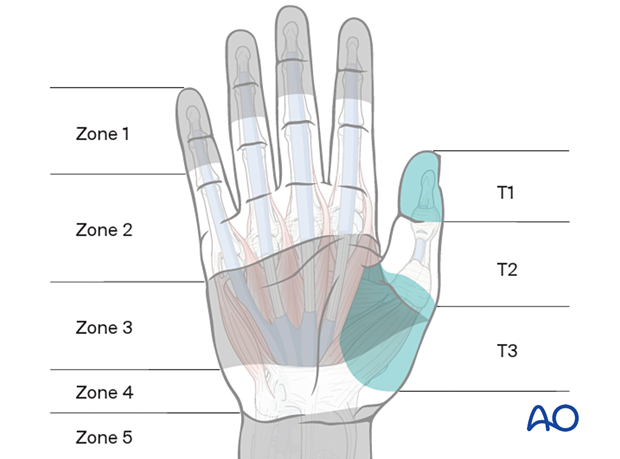
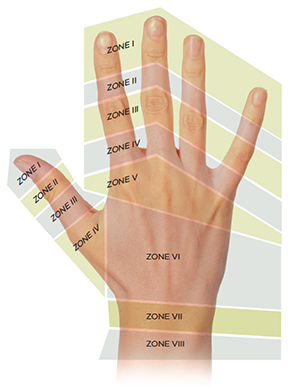
Zones of Injury
Flexor
Extensor
Basic Screening Exam:
This exam can be used in the case of a fracture/dislocation at or proximal to the wrist, or in a general trauma to ensure that there has not been a nerve injury – from the cervical spine, through the brachial plexus, and into the extremity.
Vascular
- palpate the radial pulse and check digital capillary refill.
- finger pulse oximeter is a useful adjunct for evaluating perfusion; anything below 95 in a traumatized limb/digit raises concern.
Neuro
| Nerve | Roots | Motor Test | Sensory Test |
|---|---|---|---|
| Radial | C5–C8 | Extend the wrist. If too painful due to injury, extension of the thumb IP joint may be substituted. | Dorsal webspace between the thumb and index finger |
| Median | C5–T1 | Recurrent motor branch: Opposition (thumb tip to small finger tip). Anterior interosseus branch: OK sign — touch tip of thumb to tip of index finger (note: an incorrect OK sign shows flexor substitution) | Palmar surface of the index finger or thumb |
| Ulnar | C8–T1 | Spread fingers against resistance | Palmar aspect of the little finger |
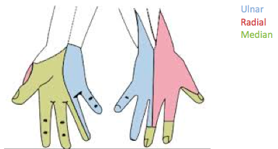
CORRECT OK SIGN
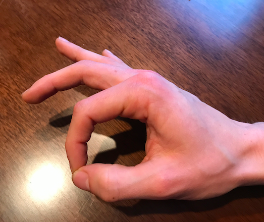
INCORRECT OK Sign
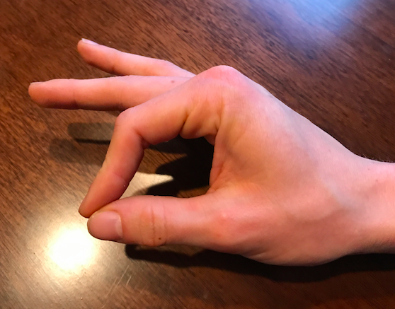
Check the individual digital sensory nerves to any finger by testing the radial and ulnar sides of each digit
If the patient can perform each of the above functions and has intact sensation, as well as good cap refill and pulses, they have passed the basic screening exam and are “neurovascularly intact.”
Detailed Hand Exam
Inspection
Look for the following findings:
Asymmetry
Lacerations/Abrasions — Any skin break over a joint (e.g., fight bite) may look innocent but provides a route for joint inoculation and can be serious.
Inflammation — May be acute (recent injury/infection) or chronic (e.g., RA).
Atrophy — Consider the distribution carefully:
- Diffuse vs. nerve-specific (e.g., thenar wasting in carpal tunnel syndrome; hypothenar + first dorsal interosseous wasting in cubital tunnel syndrome)
Traumatic Deformity — Check alignment in both flexion and extension. Abnormal rotation may only appear when making a fist.
| Finding | Description | |
|---|---|---|
| Mallet finger | Flexed DIP with inability to actively extend → rupture/avulsion of terminal extensor tendon | 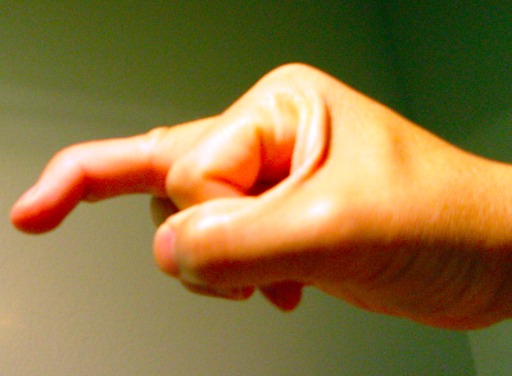 |
| Boxer’s fracture | ”Dropped knuckle sign” — metacarpal head appears to disappear due to shaft fracture | 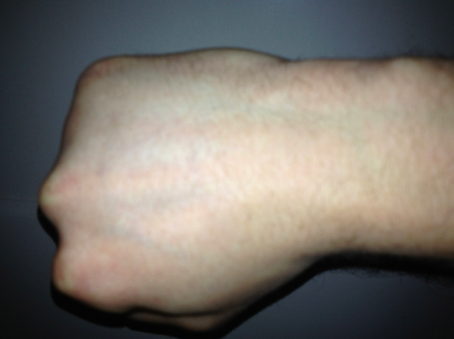 |
| Cascade disruption | One finger out of alignment with the others → may represent tendon injury | 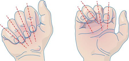 |
Palpation
Key structures to palpate, with the scaphoid being highest yield:
| Palpation Site | Landmark | Notes | Image |
|---|---|---|---|
| Scaphoid tubercle | Volar wrist | Most sensitive for scaphoid fracture (95%) | 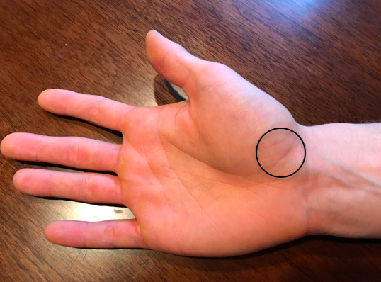 |
| Scaphoid waist | Anatomic snuffbox | 85% sensitive; maximize by ulnar deviation + thumb abduction | 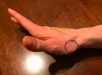 |
| Proximal scaphoid | Dorsal wrist | Soft spot between 3rd/4th extensor compartments, just distal to Lister’s tubercle | 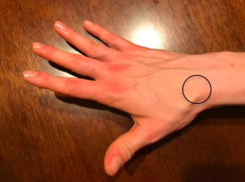 |
Range of Motion
Test both passive and active ROM in each joint.
- Passive ROM → informs about the joint itself (clicking, catching, crepitance)
- Active ROM → informs about nerve function, muscle strength, joint stability, and tendon integrity
Muscle/Tendon Exam
Volar Forearm Laceration: Finger & Wrist Flexors
Innervation: mostly median nerve, except FCU and FDP to ring/small fingers (ulnar nerve)
| Structures at Risk | Test |
|---|---|
| FPL (Flexor Pollicis Longus) | Flex thumb at IP joint |
| FDP (Flexor Digitorum Profundus) | Stabilize PIP, ask patient to flex DIP of index or middle finger |
| FDS (Flexor Digitorum Superficialis) | Hold all other fingers in extension, ask patient to flex PIP of target finger |
| FCR / FCU | Ask patient to volar-flex wrist; palpate tendon/muscle contraction |
Summary — Testing the Flexors:
- Flex thumb IP joint
- Stabilize PIP → flex each DIP in succession
- Hold remaining fingers in extension → flex each PIP in succession
- Volar (palmar) flex wrist
Dorsal Forearm Laceration: Finger & Wrist Extensors
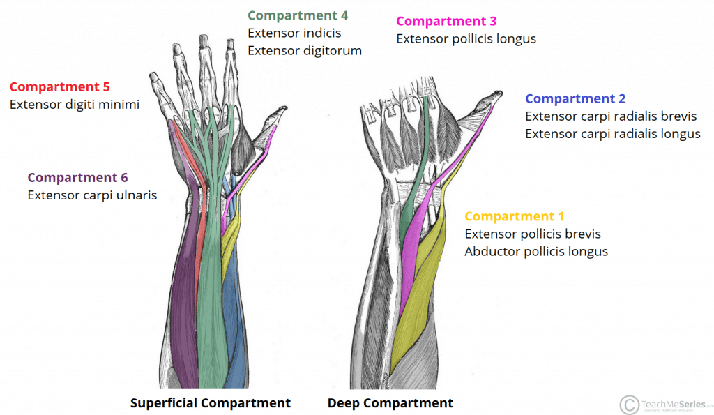
Innervation: all radial nerve, organized into 6 compartments
| Compartment | Structures at Risk | Test |
|---|---|---|
| 1st | APL, EPB | Abduct thumb; palpate tendons along radial wrist border |
| 2nd | ECRL, ECRB | Make fist, extend wrist against resistance |
| 3rd | EPL | Place hand flat on table; lift thumb off surface |
| 4th | EDC, EIP | Extend individual fingers at MCP; isolate EIP by extending index with rest of fingers in a fist |
| 5th | EDM | Extend small finger from closed fist |
| 6th | ECU | Extend and ulnar-deviate wrist |
 |
Summary — Extrinsic Extensors:
- Abduct thumb → place hand flat and lift thumb off table
- Extend fingers against resistance at MCP
- Make fist → extend wrist against resistance
- Make fist → ulnar deviate
- Extend index finger from closed fist
- Extend small finger from closed fist
Palmar Laceration: Intrinsic Muscles
Innervation: median and ulnar nerves
| Group | Muscle(s) at Risk | Nerve | Test |
|---|---|---|---|
| Thenar | APB, Opponens Pollicis, FPB | Median | Touch thumb and small finger tips together so nails are parallel |
| Thenar | Adductor Pollicis | Ulnar | Froment’s sign: hold paper between thumb base and radial index finger; weakness = thumb IP flexion to compensate |
| Lumbricals | Lumbricals | Ulnar | Flex MCP, straighten IP joints |
| Interossei | Dorsal/Palmar Interossei | Ulnar | Hand flat on table, hyperextend MCP, adduct/abduct fingers |
| Hypothenar | ADM, Opponens Digiti Minimi | Ulnar | Abduct small finger; bring small finger toward thumb |
Summary — Intrinsic Muscles:
- Touch small fingertip to thumb so nails are parallel
- Pinch paper between thumb and radial side of index finger (Froment’s sign)
- Flex MCP, straighten PIP/DIP
- Hand flat on table → hyperextend MCP → adduct/abduct each finger
- Spread fingers against resistance
Named Injuries
| Case | Diagnosis | Mechanism | Test | Example Image |
|---|---|---|---|---|
| Skier’s/Gamekeeper’s thumb | UCL rupture of thumb MCP | Fall backwards onto hand while gripping | Hold thumb metacarpal, fully extend MCP, apply radial deviation force; repeat at 30° MCP flexion. Compare to contralateral thumb. | 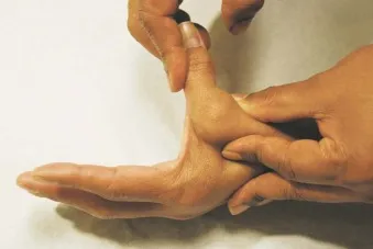 |
| [Jersey finger](Jersey Finger - Hand - Orthobullets) | FDP avulsion from distal phalanx | Finger snagged, sudden distraction force | Hold MCP and PIP in full extension → ask patient to flex DIP. Inability = positive. | 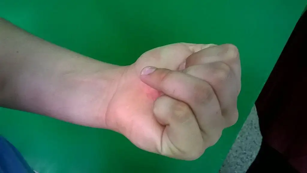 |
| Mallet finger | Extensor digitorum avulsion from distal phalanx | Axial force to fingertip (e.g., jammed on ball) | Hold middle phalanx, ask patient to actively extend DIP. Also test if patient can hold passively extended position. | 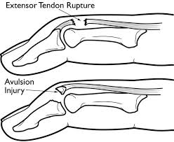 |
| Central slip injury (boutonniere deformity) | Rupture of central extensor band → lateral bands slide volar | Laceration over dorsal middle phalanx | Elson’s test: Passively flex PIP to 90°, ask patient to extend against resistance on middle phalanx. If DIP becomes taut (not floppy) → central slip injured. | 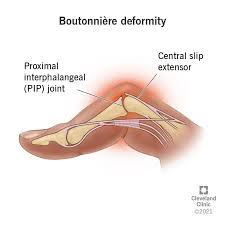 |
Pearls
- Systematic approach is essential — pain, intoxication, fear, and bleeding all degrade the exam
- A quick screening sequence: fist → open hand → wrist flexion/extension → finger extension → thumb IP flexion/extension → FDP/FDS each finger → OK sign → finger spread → gross sensation each fingertip and dorsum
- Use pulse oximetry on individual fingers to detect threatened digits before clinical ischemia is obvious; Doppler is a useful adjunct for digital vessel assessment
- Think proximal to distal — a wrist-level injury means finger flexion tests won’t tell you about the median nerve
- Edema and pain can both cause sensory changes independent of nerve injury, especially with tourniquet use
- Always document sensory exam before administering local anesthesia
References
-
[Peer-Reviewed, Web Publication] Whipple T, Gappmeier V (2018, April 16). Demystifying the Hand Exam. [NUEM Blog. Expert Commentary by Giladi A ]. Retrieved from http://www.nuemblog.com/blog/hand-exam
-
Ghane, MR et al. How trustworthy are clinical examinations and plain radiographs for diagnosis of scaphoid fractures. Trauma Mon Nov 2016. 21(5): e23345.
-
Giuglae, J et al. The palpable scaphoid surface area in various wrist positions. Journal of Hand Surgery. 1 Oct 2015. 40(1): 2039-2044.
-
Netters Orthopedic Clinical Exam. Ed: Cleland, Joshua A., PT, DPT, PhD; Koppenhaver, Shane, PT, PhD; Su, Jonathan, PT, DPT, LMT. Third Edition. Copyright 2017
-
Lin, M. Quick Tip: Elston’s Test for the Finger. Jul 29 2013. ALiEM.
-
Bookman, A. A., von Schroeder, H. P., & Pham, A. G. (2010). The Wrist and Hand. In Fam’s Muskuloskeletal Exam and Joint Injection Techniques (pp. 29–43). Mosby.
-
Seiler, JG. (2002). Essentials of Hand Surgery (pp 23-48). Lippincott Williams & Wilkins.