Carpal Bone Injuries - The Big 4 | Emergency Medicine Cases
The Big 4 carpal bone injuries
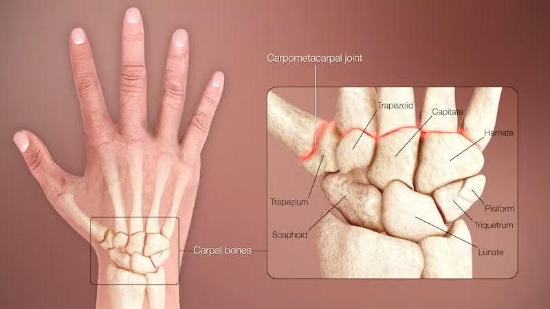
The following carpal bone injures are commonly missed with serious consequences for patients. Other carpal bone injuries are less common and may co-occur with the “Big 4” injuries:
- Triquetrum chip fractures
- Scapholunate sprains/dissociation/dislocation
- Hook of the hamate fracture
- Scaphoid fracture
General Principles
- Age matters: Pediatric and elderly patients → more likely distal radius fractures after FOOSH. Carpal bone injuries occur almost exclusively in young adults.
- Volar vs. dorsal injuries:
| Type | Mechanism | Stability | ED Management |
|---|---|---|---|
| Dorsal (e.g., Colles) | FOOSH | More stable | Below elbow splint often adequate |
| Volar (e.g., Smith’s, volar Barton’s) | Fall on back of hand | More unstable | Urgent ortho involvement; above elbow splint |
- Complete the physical exam before ordering x-rays — helps narrow differential and guide imaging.
- Always test pronation/supination — limitation raises suspicion for DRUJ (Distal Radius-Ulna Joint) injury.
Physical Exam: Surface Anatomy
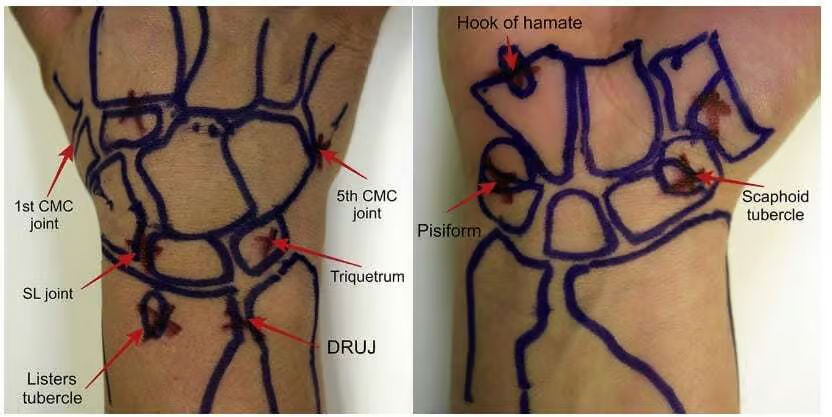
Dorsal Wrist — 4 Key Divots
| Structure | Location |
|---|---|
| Scapholunate (SL) space | Divot just distal to Lister’s tubercle |
| Triquetrum | Divot just distal to ulnar styloid |
| Scaphoid snuffbox | Palpate in ulnar deviation; compare to contralateral side |
| DRUJ | Divot between distal radius and ulnar styloid |
Volar Wrist — 3 Key Bumps
| Structure | Location |
|---|---|
| Scaphoid tubercle | Just proximal to thenar eminence base; start in ulnar deviation, move to radial deviation |
| Pisiform | Only volar carpal bone; just proximal to hypothenar eminence base |
| Hook of hamate | 2cm distal and 1cm radial to the pisiform; requires deep palpation |
⚠️ Pitfall: Don’t skip pronation/supination ROM — subtle DRUJ injuries will be missed.
X-Ray Interpretation
Standard Views
AP, lateral, and oblique. The lateral is the primary view — most commonly reveals subtle missed injuries. View the lateral with the forearm in the horizontal plane, thumb pointing down.
AP View — 2 Key Alignment Patterns
| Finding | Normal | Abnormal Suggests |
|---|---|---|
| Distal radius-ulna space | ≤ 2mm | > 2mm → DRUJ dislocation |
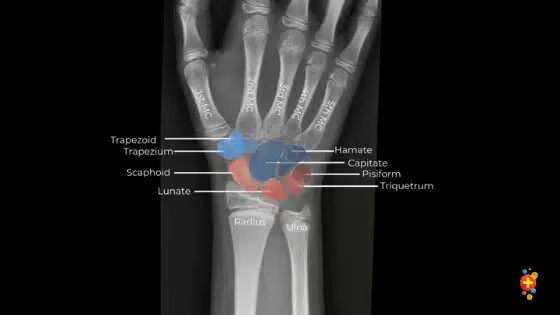
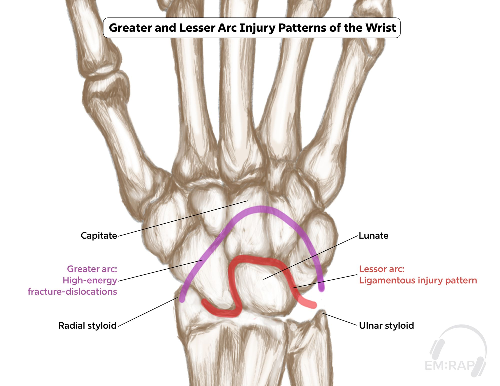
| Gilula’s 3 arcs | Smooth arcs, equal carpal spaces | Disruption → carpal bone injury |
Lateral View — 2 Key Alignment Patterns
| Finding | Normal | Abnormal Suggests |
|---|---|---|
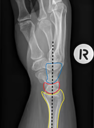
| Radius-lunate-capitate alignment | Collinear | Malalignment → scapholunate injury |
| Radius-ulna overlap | Present | Absent → DRUJ injury |
 |
Bone Shapes to Know
- Scaphoid on AP → normally boat-shaped
- Lunate on lateral → half-moon; on AP → roughly square
X-Ray Signs of Lunate/Perilunate Pathology
| Sign | View | Appearance | Diagnosis |
|---|---|---|---|
| Signet ring sign | AP | Rounded cortex of scaphoid tubercle | Scapholunate subluxation (scaphoid flexion) |
| Piece of pie / pizza sign | AP | Triangular appearance of lunate | Lunate dislocation |
| Empty teacup sign | Lateral | Capitate no longer sits in lunate concavity | Perilunate dislocation |
DRUJ Injuries: “The Forgotten Joint”
Why It Matters
A missed DRUJ injury can lead to chronic supination deficit and pain — preventable with early surgery in some cases.
Mechanism
Forced supination/pronation: power tools, fall while carrying heavy load, MVC with tight grip on steering wheel. Also any FOOSH.
Physical Exam
| Maneuver | Finding |
|---|---|
| Active/passive supination-pronation against resistance | Pain |
| Point tenderness over DRUJ | Present |
| DRUJ shuck/ballottement test | Grasp ulnar styloid, stabilize distal radius → increased mobility vs. contralateral side |
| Piano key sign | Ulnar styloid appears protuberant, easily ballottable, rebounds to dislocated position after volar depression |
X-Ray Findings
- AP: DRUJ widening > 2mm
- Lateral: loss of radius-ulna overlap
- Often occult on x-ray — exam is key
Associated Fractures
- Any distal radius fracture (all displaced distal radius fractures have some degree of DRUJ injury)
- Galeazzi fracture: dorsally angulated distal 1/3 radius fracture + DRUJ dislocation → usually requires ORIF
- Radial neck fracture
ED Management
Immobilize in supination to block pronation:
- Deep radial gutter splint
- Above elbow back slab
⚠️ Pitfall: Do NOT use a short below-elbow splint for DRUJ injuries — always above elbow with forearm supinated.
Distal Radius Fractures
Immobilization Principles
| Fracture Type | Mechanism | Splint Position | Molding? |
|---|---|---|---|
| Colles (dorsal) | FOOSH | Flexion + ulnar deviation | ✅ Yes |
| Smith’s (volar) | Fall on back of hand | Extension + radial deviation | ❌ No — risk of median nerve compression |
| Volar Barton’s | Fall on back of hand | Above elbow splint; early ortho | ❌ No |
| Dorsal Barton’s | FOOSH variant | Often surgical | — |
⚠️ Reduce in the opposite direction of the injury mechanism.
Barton Fractures — Don’t Be Fooled
Both Barton variants may appear minor on x-ray but are often unstable and require surgery:
- Dorsal Barton’s — fracture of dorsal lip of distal radius extending into joint; associated radiocarpal subluxation/dislocation; distinguish from Colles as management is more often surgical
- Volar Barton’s (reverse Barton) — fracture of volar lip; unstable; requires ORIF
Triangular Fibrocartilage Complex (TFCC) Injury
Think of the TFCC as “the meniscus of the wrist.” Displaced distal radius fractures may be associated with a TFCC tear, which typically causes chronic ulnar-sided wrist pain after the fracture has healed. Not usually diagnosed acutely in the ED, but worth noting in discharge counseling and ortho follow up planning.