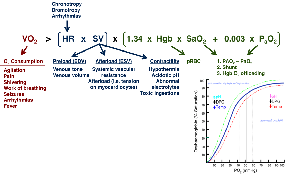
Oxygen Delivery (DO2) = Arterial O2 Content (CaO2) x Cardiac Output (CO)
DO2 = CO x (Hgb x SaO2 x 1.34 + PaO2 x 0.003)
Arterial Oxygen Content (CaO2)
Arterial Oxygen Content (CaO2)
- CaO2 = (Hgb x SaO2 x 1.34) + (PaO2 x 0.003)
- Hgb: Hemoglobin
- Oxygen Carrying Capacity = 1.34
- SaO2: Percent Arterial Oxygen Saturation of Hgb
- SpO2: Peripheral Oxygen Saturation is Detected on Pulse Oximeter and Used to Estimate SaO2
- PaO2: Partial Pressure of Dissolved Oxygen in Blood (Oxygen Tension)
Level of Importance
- Hemoglobin is Generally the Most Clinically Significant Determinant of Arterial Oxygen Content (CaO2)
- SaO2 Has the Same Impact as Hemoglobin but Generally Has Less Variance
- PaO2 is the Least Important Measure and Only Contributes 1-2%
Cardiac Output (CO)
Cardiac Output (CO)
- CO = Heart Rate (HR) x Stroke Volume (SV)
- Factors that Increase Stroke Volume:
- Increased Preload (End-Diastolic Volume)
- Increased Contractility
- Decreased Afterload
Oxy-Hgb Dissociation Curve
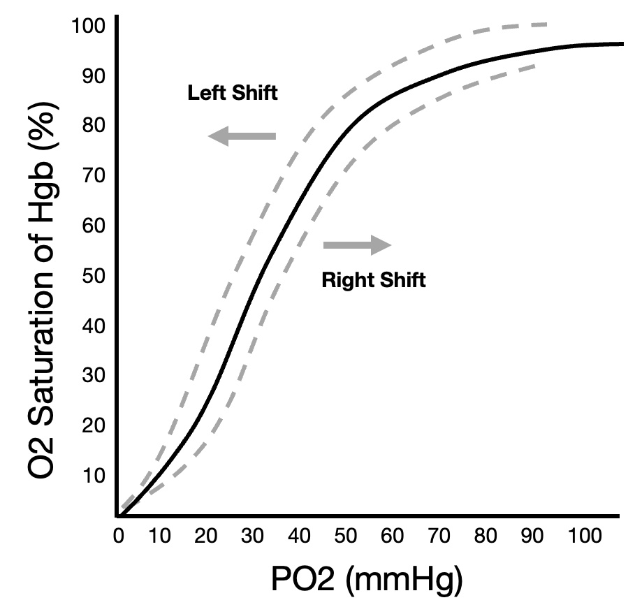
Compares Partial Pressure of Oxygen (PaO2) to Percent Oxygen Saturation of Hemoglobin (SaO2)
- Shifts:
- Left Shift – Increased Affinity Supports O2 Binding
- Right Shift – Decreased Affinity Causes O2 Unloading
- Factors that Cause a Leftward Shift:
- Decreased CO2
- Decreased Temperature
- Decreased H+ (Increased pH)
- Decreased 2,3-Diphosphoglycerate (DPG)
- Carbon Monoxide Exposure – Increases Affinity at Other Binding Sites
- Factors that Cause a Rightward Shift:
- Increased CO2
- Increased Temperature
- Increased H+ (Decreased pH)
- Increased 2,3-DPG (2,3-Diphosphoglycerate)
- Hypoxia Further Decreases Oxygen Affinity Due to 2,3-DPG Having Increased Affinity for Deoxygenated Hemoglobin
- Fetal Hemoglobin Has a Low Affinity for 2,3-DPG Resulting in Overall Higher Affinity for Oxygen