Quick Reference
| First 10 Minutes | |
|---|---|
| Initial Eval | - Finger stick glucose or empiric D50 ampule - Rapid neuro exam (GCS & NIHSS) - Avoid hypotension & hypoxia; Early airway management if needed - Resuscitation goal = get to CT scanner |
| BP Goal | SBP <160 or MAP <110 - Preferred antihypertensives: Nicardipine, oral Nimodipine; - Consider arterial line for precise monitoring |
| Labs | - CBC / CMP - Coags (if on anticoagulation or unknown) |
| Imaging | CT Head w/o contrast + CTA vs LP if >6 hours from onset |
| Consults | Neurosurgery |
Ottawa Subarachnoid Hemorrhage (SAH) Rule for Headache Evaluation
- 100% sensitive / 15% specific
Imaging Reference: The Radiology Assistant : Non-traumatic Intracranial Hemorrhage
Evaluation
Think about SAH in patients presenting with sudden headache PLUS:
- Altered LOC (ie. persistent GCS <15)
- Seizure
- Nausea / Vomiting
- LOC/syncope
- Neck pain (unilateral)
- Focal neurological deficits
- Low grade fever (from meningeal irritation due to the subarachnoid blood)
History
Questions
- When did you first notice the headache?
- What were you doing at that time?
- How severe was it when you first noticed it on a scale of 0-10?
- Did it progress or get worse after that?
- When did the headache reach maximal intensity?
Data
- “Thunderclap” headache – 83% of patients with SAH
- “Worst headache of life” ie. severe instantly peaking headache – 95% of patients with SAH
- BUT- 70% of patients presenting to the ED with headache but without SAH will also endorse having “worst headache of life.”
- Headache taking more than 1 hour to reach peak severity decreases the chance of patient having SAH, LR- 0.06
- Neck stiffness – 74% of patients with SAH
- Subjective neck stiffness/ pain on history, LR+ 4.1
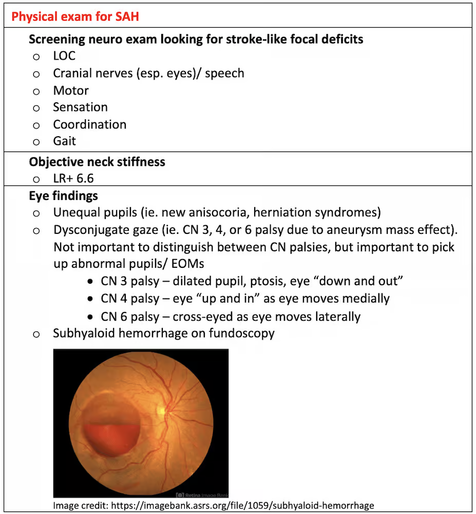
- Objective neck stiffness on exam, LR+ 6.6
- Vomiting – 62% of patients with SAH
- Loss of consciousness/ syncope – 13% of patients with SAH
Physical Exam
Workup
Initial Imaging
- CT Non-Con
- follow 6 hour rule: CTA vs LP
6 Hour Rule
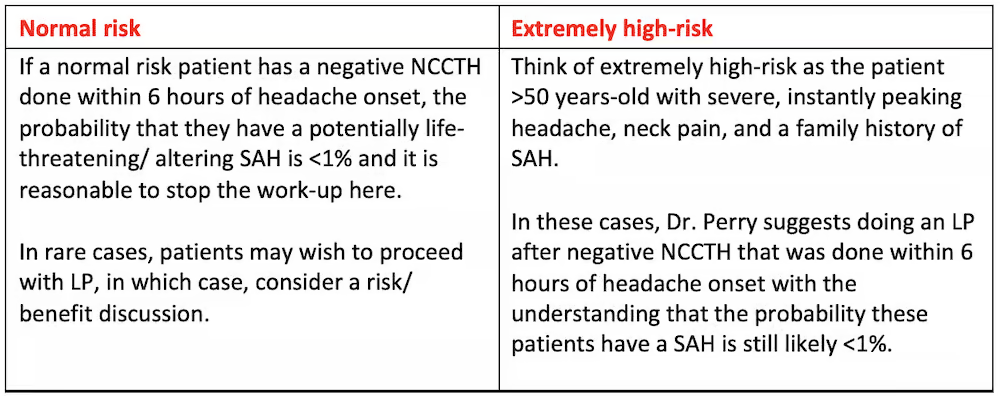
- If the patient presents <6 hours after headache onset and certain criteria are met, non-contrast head CT is diagnostic
- If the patient presents >6 hours from headache onset, non-contrast head CT is negative, and patient has a high pretest probability, then LP or CT angiography (CTA) is recommended.
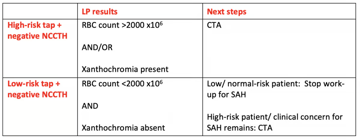
Lumbar Puncture
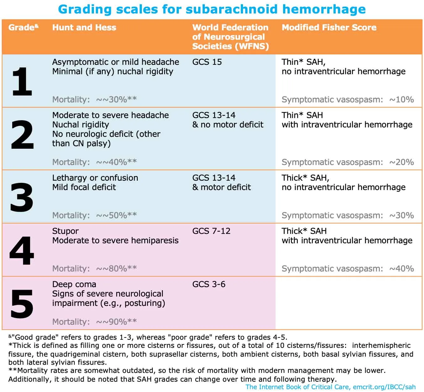
Grading
Treatment/Management
Pharm
| Class | Meds |
|---|---|
| Hyperosmolar Therapy | - hypertonic saline (3ml/kg) |
| Antiemetics: | - ondansetron - prochlorperazine |
| Analgesics: | - fentanyl - acetaminophen - gabapentin - steroids |
| Antihypertensives: | - Nicardipine IV - Nimodipine PO |
| Seizure Prophylaxis | - Levetiracetam |
| Antipyretics | - acetaminophen? |
Non-Pharm
- Head of bed at 30 degrees
- If intubated: gentle hyperventilation (pCO2 30-35)
- no more than 1-2 hours; temporizing
Procedures
- EVD
Disposition
Admit!
Full Overview
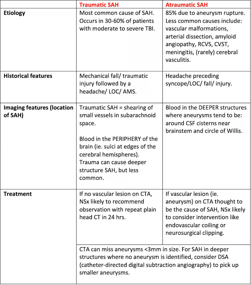
Etiology
- Primary SAH:
- 85% are aneurysmal.
- 10% are perimesencephalic hemorrhages.
- 5% are due to other vascular malformations:
- AVMs (arteriovenous malformations).
- dAVF (dural arteriovenous fistula).
- Arterial dissection.
- Moyamoya disease.
- Secondary SAH
- Trauma.
- RCVS (reversible cerebral vasoconstriction syndrome).
- PRES (posterior reversible encephalopathy syndrome).
- CVT (cerebral venous sinus thrombosis).
- CAA (cerebral amyloid angiopathy).
- Pituitary apoplexy.
- CNS vasculitis.
- Primary intracerebral hemorrhage with secondary extension to the subarachnoid space.
- Tumor.
- Coagulopathy.
- Sickle cell disease.
- Sympathomimetic abuse.
- Mycotic aneurysm (usually due to septic emboli from endocarditis; most often at distal MCA or vertebrobasilar system).(Torbey, 2019)
- Vertebral artery dissection that extends intracranially.
- Iatrogenic injury to cerebral vasculature.
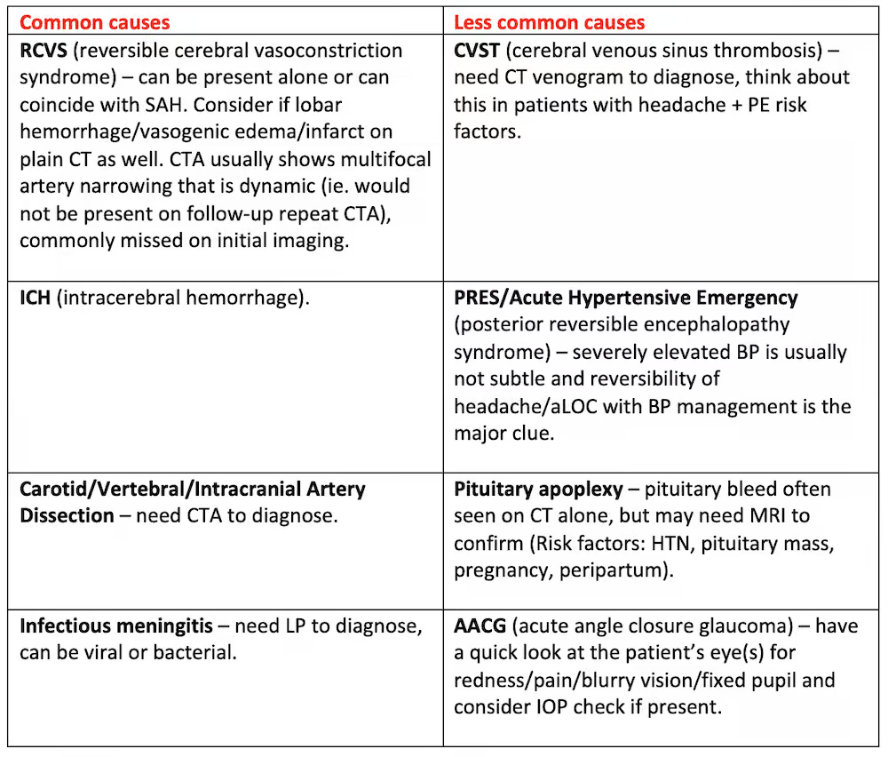
DDx for “Thunderclap Headache”
Risk Based Eval: Imaging/LP
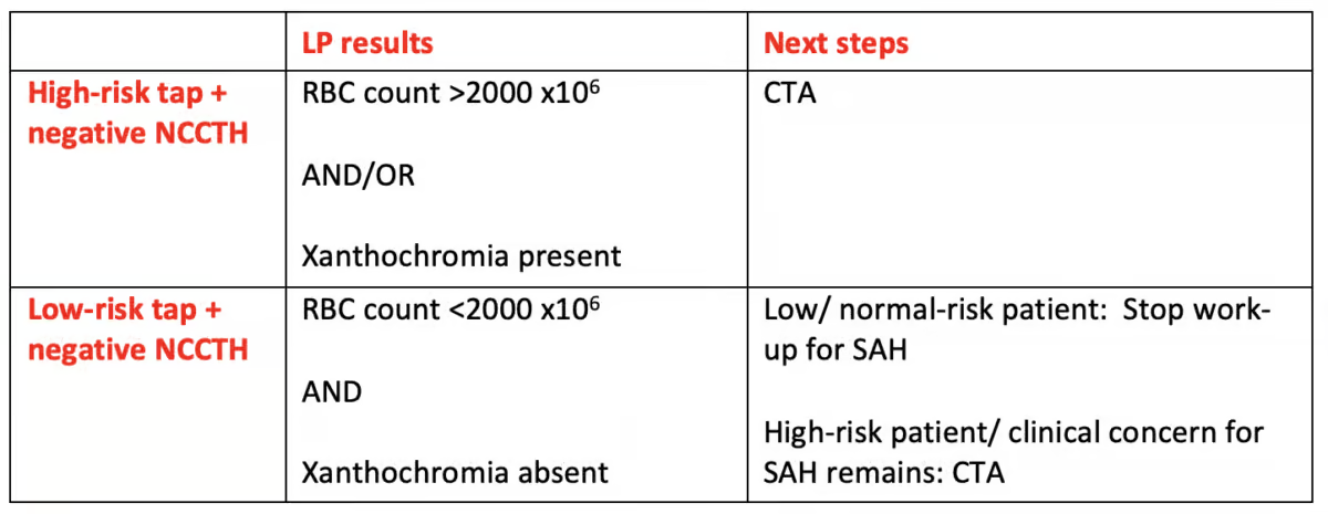
6 Hour Rule Specifics
Under the below conditions, non-contrast head CT at <6 hours has a sensitivity of 98.7% (confidence interval of 97.1%-99.4%).
Patient characteristics
- Isolated thunderclap headache
- No seizures, LOC, or neck pain
- Hematocrit (Hct) >30%
- Accurate time of onset
- Normal neurologic examination and no meningismus
Test characteristics: - Indication listed as “rule out aneurysmal subarachnoid hemorrhage or thunderclap headache”
- Third-generation CT scanner or newer
- Image is technically adequate (no motion artifact)
- Thin cuts ≤5mm through base of brain
- Hct >30%
- Attending level radiologist who routinely reports on head CT images
CTA
- Performance: highly sensitive (~97%) and specific (~95%) for aneurysm detection. Very small aneurysms may be missed.
- CTA has numerous potential roles:
- (1) For patients who have a SAH, CTA is essential to evaluate for underlying vascular anomalies (e.g., aneurysms, arteriovenous malformations).
- (2) For patients who do not have a SAH, CTA is useful to evaluate for the possibility of Reversible Cerebral Vasoconstriction Syndrome (RCVS) or cervical artery dissection.
- (3) For patients with a thunderclap headache and possible SAH, the finding of an aneurysm may indicate the need for further diagnostic testing (e.g., with lumbar puncture).
- Given the increasing recognition of RCVS as a cause of thunderclap headache, patients with thunderclap headache may benefit from CTA, regardless of whether the noncontrast CT is positive for SAH.
- Broader use of CTA may also be bolstered by evidence that contrast dye is not nephrotoxic.
- A potential drawback of CTA is that it may reveal asymptomatic unruptured aneurysms in ~2% of the population.
LP
Looking for: (1) RBCs and/or (2) xanthochromia.
***Dont wait for 12 hours to pass
- RBCs: Intact RBCs will be seen early in the course of aneurysmal subarachnoid hemorrhage before lysis occurs in the CSF. Because of the occasional difficulties experienced in completing LP, RBCs can sometimes be seen in CSF tubes from local tissue trauma, termed a “traumatic tap.”
- If the CSF contains RBCs, the likelihood of aneurysmal subarachnoid hemorrhage (vs. traumatic tap) increases with higher RBC counts. Many use a cut-off point of >2,000 × 106/L RBCs in the fourth tube to distinguish an aneurysmal subarachnoid hemorrhage from a traumatic tap.
- Note that this cut-off point does not exclude all instances of traumatic tap but significantly decreases the likelihood of traumatic tap and improves the LP test characteristics. It is not, however, a universally agreed upon cut-off.
- Xanthochromia: RBC lysis in the CSF results in a yellow discoloration. This process can take time to occur, and traditional teaching has stated that the LP should be performed >12 hours from ictus, when xanthochromia is consistently expected.
Risk Factors
- Standard vascular risk factors: hypertension, smoking, age >50
- Personal history of cerebral aneurysm: especially if >5mm in size, any documented growth on serial imaging, and/or located in the posterior circulation vessels
- Genetic risk factors: family history of cerebral aneurysm or aneurysmal SAH, connective tissue diseases like Elhers-Danlos (less so Marfan Syndrome), autosomal dominant Polycystic Kidney Disease
- High-risk substance use: binge or chronic ETOH, sympathomimetic use (cocaine, methamphetamines)
References
Writeup and Original Sources contained within:
Subarachnoid Hemorrhage Recognition, Workup & Diagnosis | EM Cases
Management of Subarachnoid Hemorrhage | Emergency Medicine Cases
Subarachnoid Hemorrhage (SAH) - EMCrit Project
Subarachnoid Hemorrhage: Thunderclap Headache - EM Board Bombs Podcast