Quick reference
2026 Guidelines
Evaluation
EKG
- [PE EKG Findings](Is this ECG easy for you yet? - Dr. Smith’s ECG Blog)
- EMCrit Wee - ECGs in Acute Pulmonary Embolism - Death Threats in Little Squiggly Lines
Simultaneous T-Wave Inversions in V1-4 AND III/AVF (anterior and inferior leads)
- Pattern: little R, big S, coved ST, bit TWI
- Sinus Tachycardia
- S1Q3T3
- Ultrasound (McConnell’s, D sign, etc)
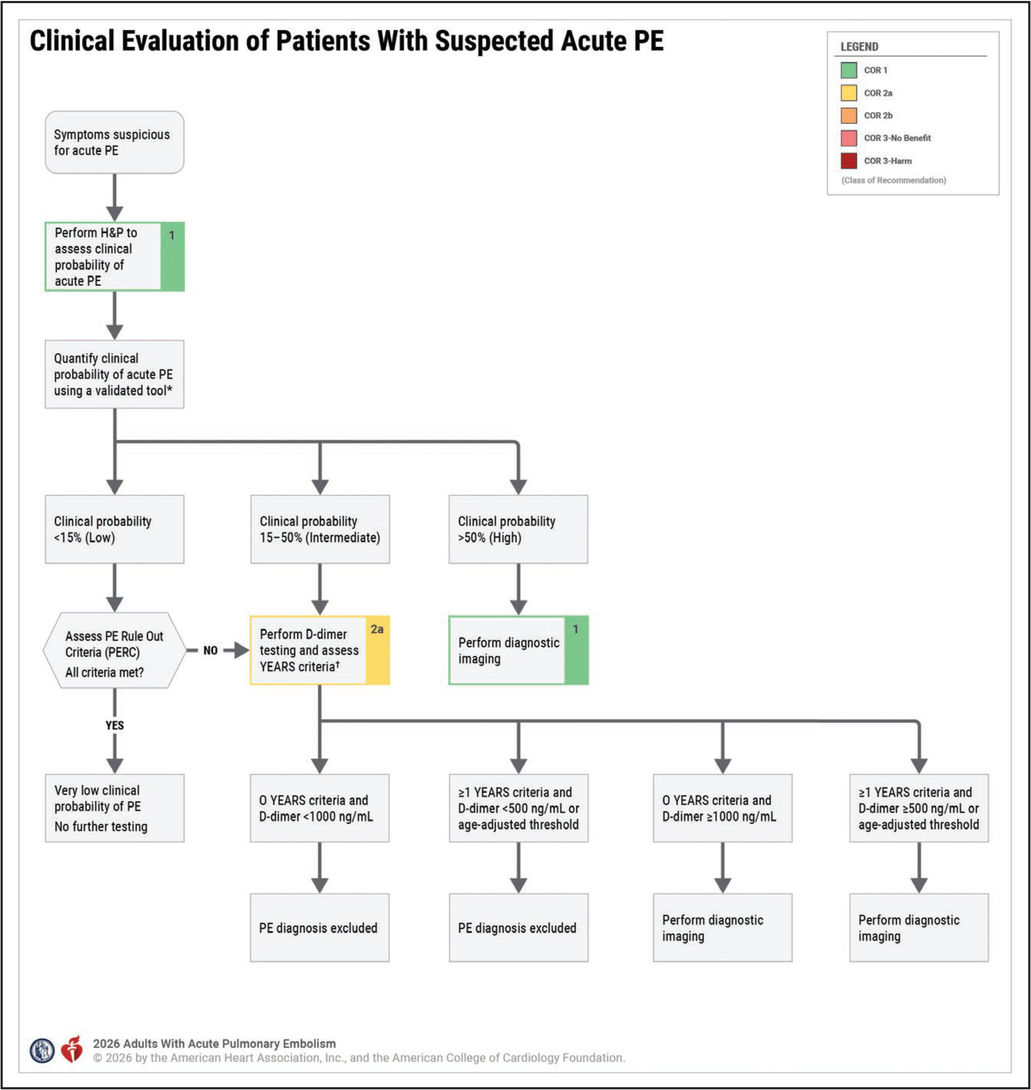
Scores
Well’s
- Low Risk: PERC
- Intermediate: D-Dimer / YEARS
- High: CTPA
Wells’ Criteria for Pulmonary Embolism
PERC Rule for Pulmonary Embolism
YEARS Algorithm for Pulmonary Embolism (PE)
Age-Adjusted D-dimer for Venous Thromboembolism (VTE)
History and Physical Exam
Symptoms:
Especially concerning:
Tachycardia + hypotension = likely RV dysfunction
Tachypnea + hypoxemia = significantly impaired VQ
- one of four key symptoms are present in 94% of patients
- sudden onset dyspnea
- pleuritic chest pain (especially concerning when localizable)
- syncope
- hemoptysis
- Tachycardia (heart rate >100/min) occurs in 65-70% of PE patients
- Hemoptysis, though present in only 5-13% of patients with PE, is a particularly specific finding
- Clinical signs of DVT (including unilateral leg swelling, tenderness, and prominent collateral veins) are present in approximately 40% of symptomatic PE patients and substantially raise the probability of diagnosis.
Risk factors:
- Recent surgical procedures (especially orthopedic).
- Recent hospitalization for heart failure or MI.
- Prior venous thromboembolic disease.
- Malignancy (especially metastatic).
- Postpartum (especially after C-section).
- Estrogen-containing oral contraceptives, hormone replacement therapy, in vitro fertilization.
- Older age.
- Immobility.
- Thrombophilia (including autoimmune diseases, inflammatory bowel disease, infection).
- Approach to PE diagnosis: (https://emcrit.org/ibcc/pulmonary-embolism/#approach_to_PE_diagnosis)
PE Severity
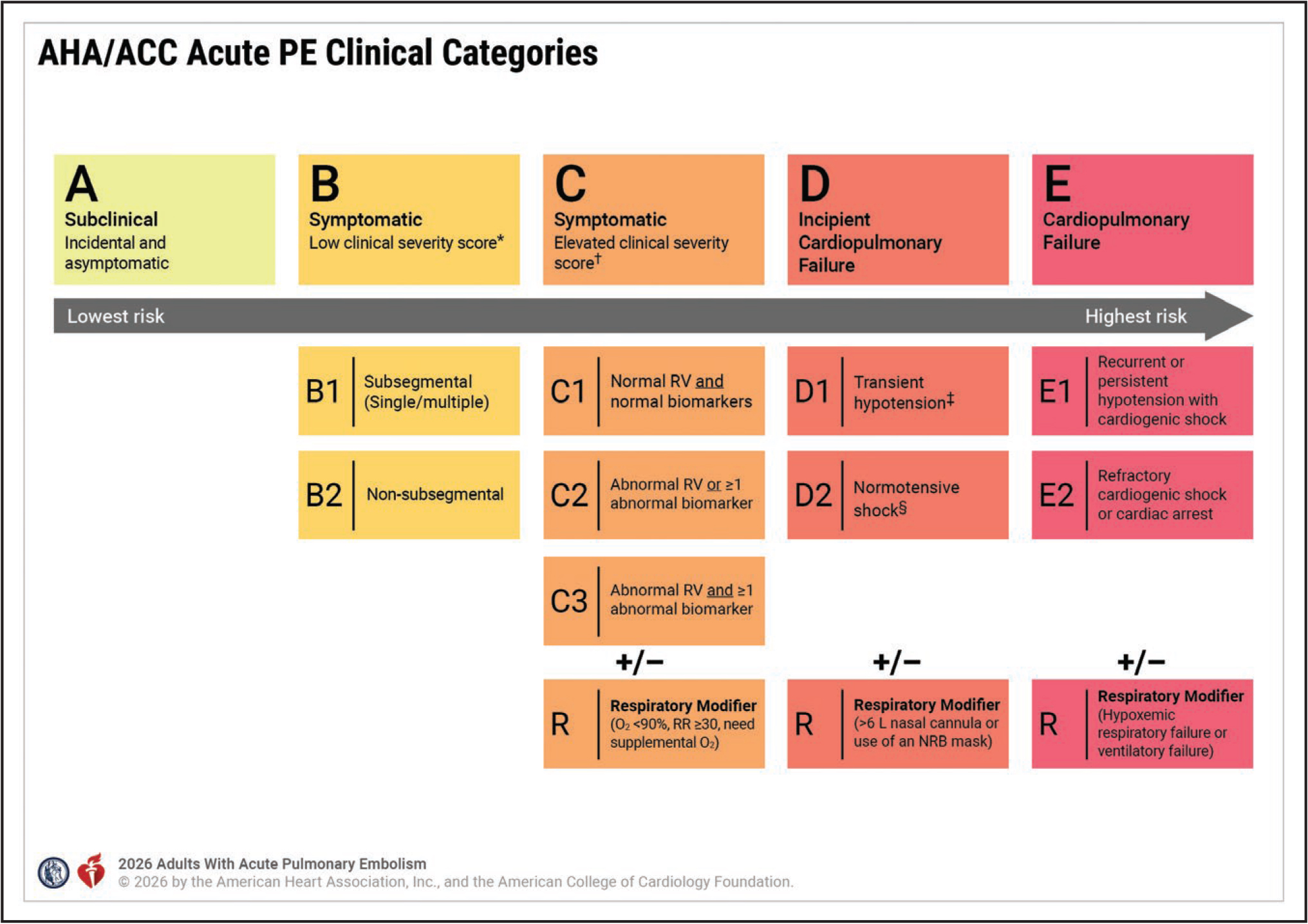
Additional Initial Workup by Clinical Category
Determine Severity
Pulmonary Embolism Severity Index (PESI)
Simplified PESI (Pulmonary Embolism Severity Index)
- Category A / B:
- severity score (PESI / sPESI)
- Category C / D:
- RV imaging (CT or TTE)
- cardiac biomarker (troponin)
- lactate
Treatment
Anticoagulation: All Categories
- A: DOAC
- C/D/E1: LMWH
- E2: LMHW / UFH
Advanced Therapies: D1 / D2 / E2
- Thrombectomy
- D1/D2/E1: MT acceptable
- Systemic Thrombolysis
- D/E: TPA
- Supportive / Adjunctive Care
- VA-ECMO: E1/2
Duration:
-
Provoked/Reversible risk factors: 3-6 months
- Unprovoked / irreversible risk factors: prolonged therapy
Disposition
A: outpatient treatment with DOAC
B: sPESI/PESI - Outpt / CDU (obs)
C:
- C1: floor
- C2/3: IMC
D/E: ICU
Special Populations
Pregnancy:
- Use LMWH
- AVOID DOACs and VKAs
- Low-dose CTPA > VQ scan
- VQ SPECT > Planar VQ
Breastfeeding:
- Use LMWH
- AVOID DOACs
Obesity
- DOACs preferred
CKD:
- CrCl 30-89: DOACs safe
- CrCl <15: Apixaban?
Hepatic Impairment
- Child-Pugh A/B: DOACs safe
- Child-Pugh C: Avoid DOACs
APL:
- VKAs preferred (especially for “triple positive” patients)
Brain Tumors
- DOACs preferred