Pearls
- need for rate treatment is determined by VENTRICULAR rate
- best lead for P-waves: V1
- A-fib w/ WPW: ONLY shock or procainamide (rate control will KILL)
- AIVR = common reperfusion rhythm s/p PCI/lytics in ACS
- NOT dangerous - can last for a few minutes
- Don’t treat
- V-fib: CHECK FOR PULSE
- Short PR DDX: junctional rhythm, pre-excitation syndrome
rhythms
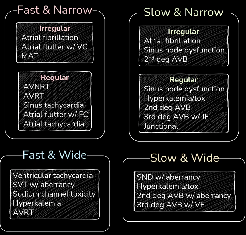
| Tachy Category | Potential Rhythms |
|---|---|
| Narrow Regular Tachycardia | Sinus Tach / SVT / A-Flutter |
| Narrow Irregular Tachycardia | A-Fib / A-Flutter w/ variable conduction (F-waves) / MAT (think COPD) |
| Wide Regular Tachycardia | Treat all as V-Tach |
| Wide Irregular Tachycardia | A-Fib w/ BBB; A-Fib w/ pre-excitation (e.g. WPW) — rates >200, irregularly wide/narrow, tx: Procainamide OR shock |
| Monomorphic Ventricular Tachycardia | Stable? → Amiodarone; Unstable? → Synchronized cardioversion. AIVR = ventricular rhythm 40–130 bpm (seen post-arrest) |
| Polymorphic Ventricular Tachycardia | Long QT → Torsades de Pointes → IV Mg!! |
DDx for Various Findings
Anterior T-wave Inversion
- abnormal depolarization (make sure it’s not the QRS’ fault - abnormal depolarization = abnormal repolarization)
- Anterior reperfusion (i.e. Wellen’s syndrome)
- Acute Right Heart Strain (PE, etc)
- Hypokalemia
- Takotsubo Cardiomyopathy
OMI / NOMI
NSTE ACS Criteria
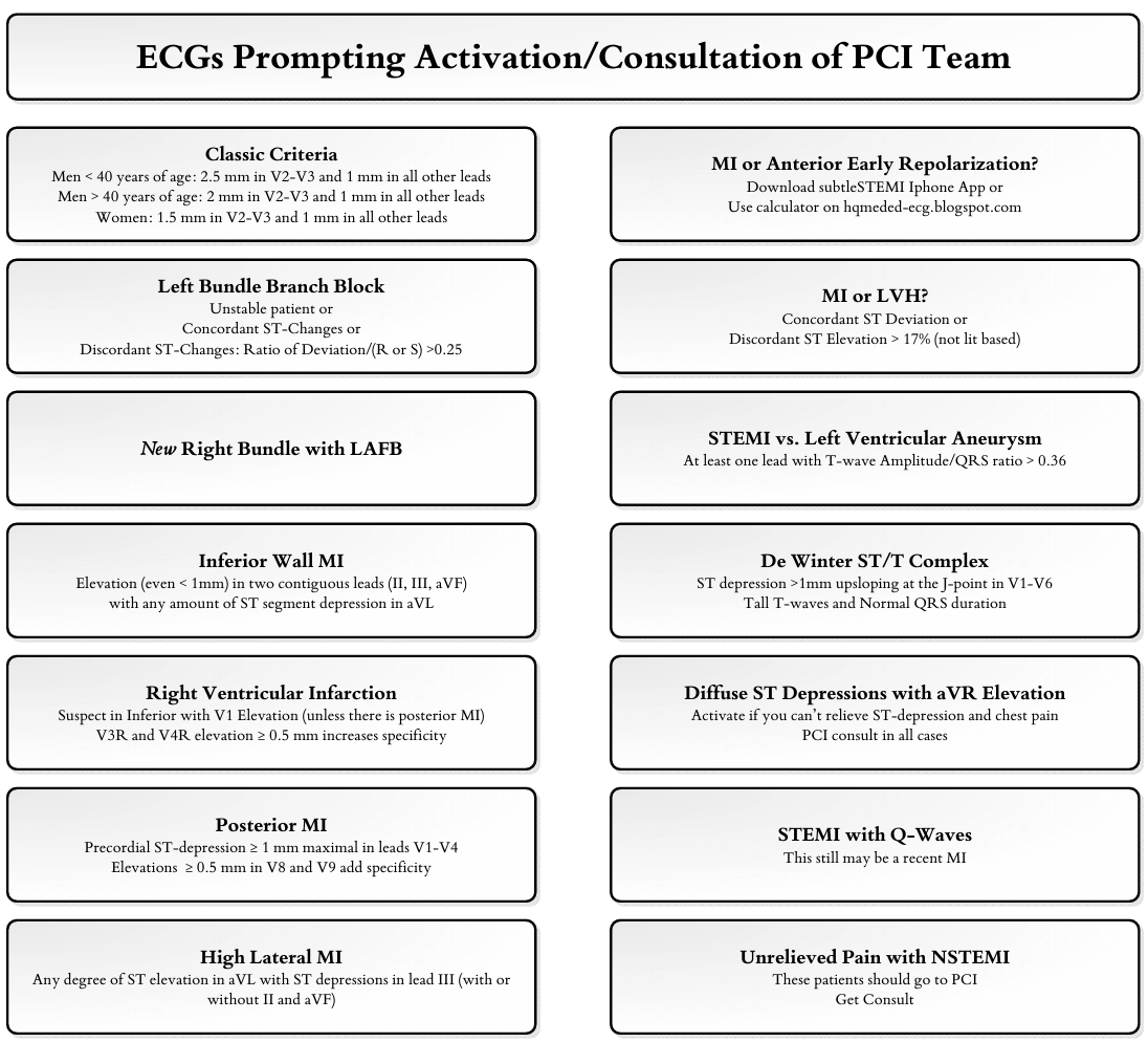
OMI needing reperfusion (STEMI and STEMI Equivalents)
Reading EKGs
EKG Overview
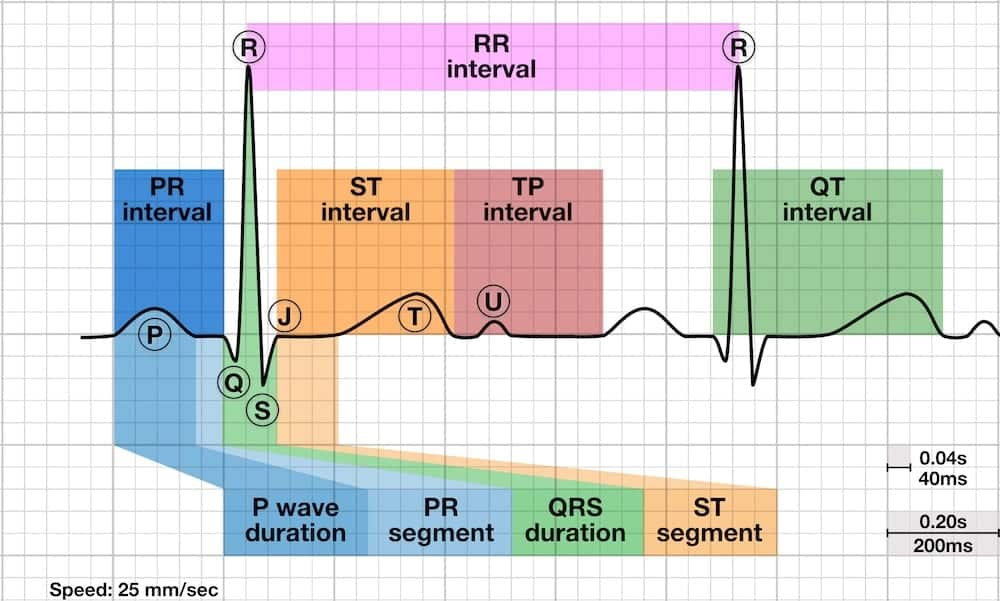
Interval Time Standards:
- 1 small box = 40ms (.04 sec)
- 1 big box = 200ms (.2 sec)
- PR- 3-5 small boxes (<1 big box)
- QRS- <3 small boxes (100-120ms)
- QT- <440ms (460ms in female)
EKG Interpretation:
note- beware of Limb Lead Reversal
- negative P wave and negative QRS complex in lead I the likely diagnosis is limb lead reversal.
1. Calculate Rate
- 300 - 150 - 100 - 75 - 60 - 50
2. Assess Rhythm
- Is it regular or irregular?
- If irregular –
- Is there a pattern or is it irregularly irregular?
- Are there premature or escape beats? Based on morphology - what type?
- every other = bigeminy
- every third = trigeminy- Are P waves present?
- Are they upright/normal in Lead II?
- Are they similar?
- Check P:QRS ratio – if not 1:1, check for AV blocks
- Are P waves present?
3. Determine axis
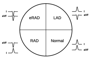
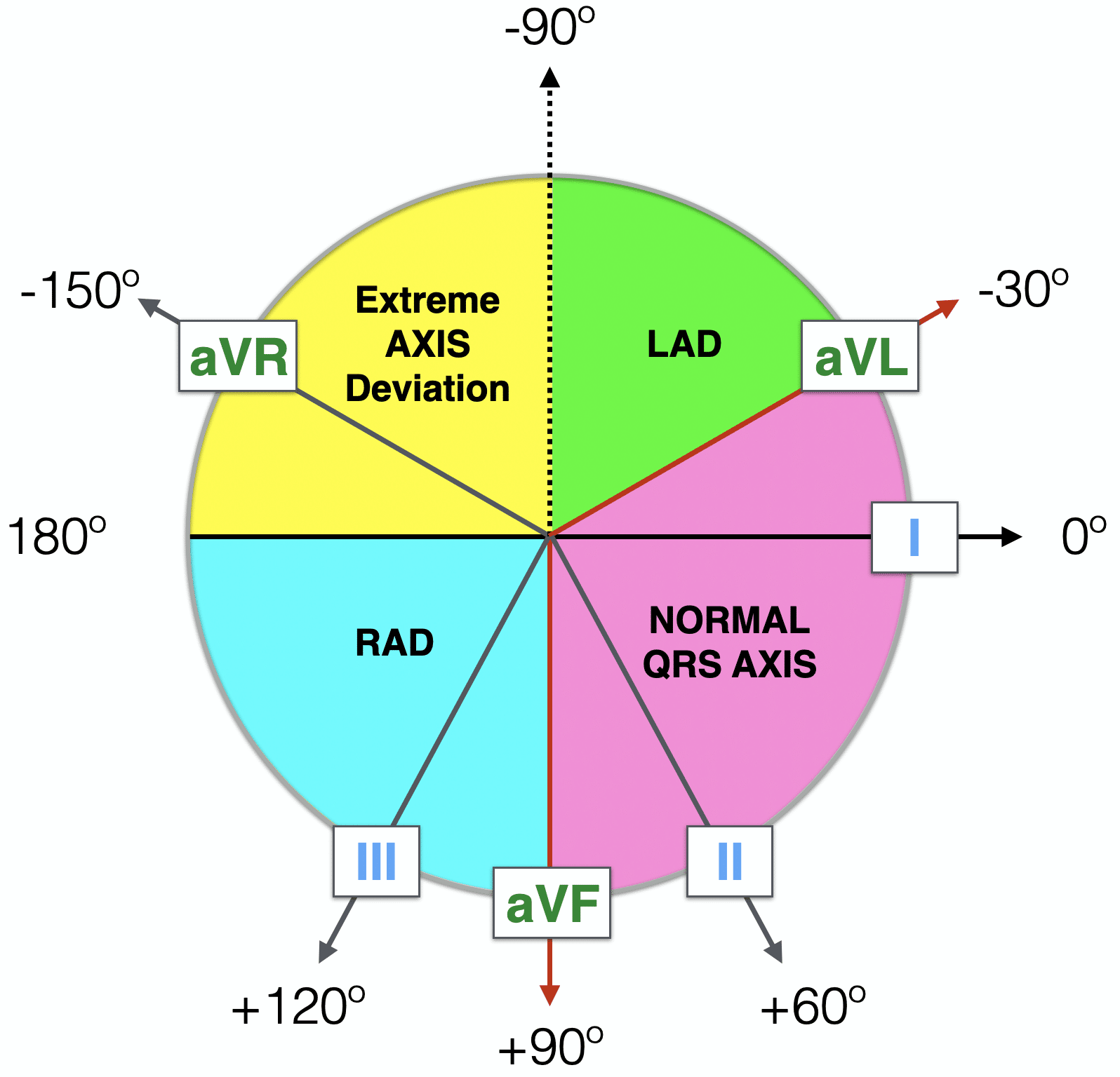
- Use Leads I and aVF to identify quadrant
- Normal – positive in Leads I and aVF
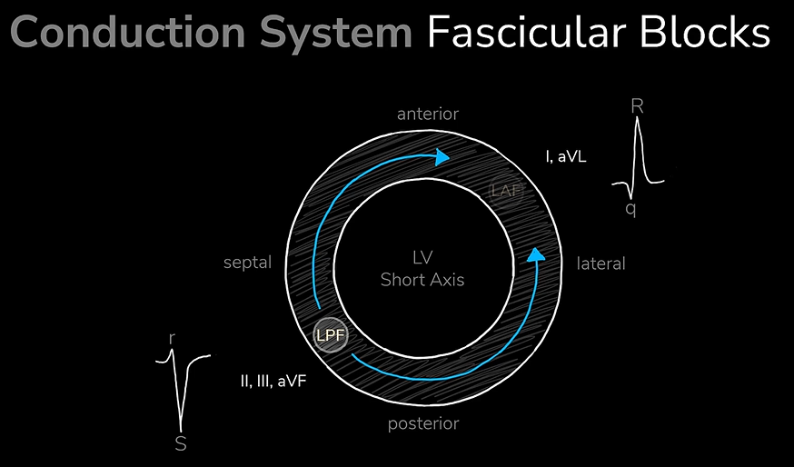
- LAD – positive Lead I, negative Lead aVF
- left bundle branch block / paced
- inferior MI
- LVH
- WPW
- LAFB
- LAD + really downgoing lead II = likely LAFB
- Dominant S in II/III/AVF and dominant R in I, AVL
- RAD – negative Lead I, positive Lead aVF
- ventricular ectopy
- lateral MI
- RVH
- WPW
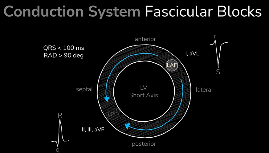
- left posterior fascicular block
- Dominant R in II/III/AVF and dominant S in I, AVL
- Associated with RBBB
- Hyperkalemia
- sodium channel toxicity (classically TCA overdose)
- Pulmonary embolism (dyspnea + RAD!!)
- COPD / chronic lung disease
- normal in children
- Extreme RAD – negative in Leads I and aVF
- Axis lies 90 degrees to most biphasic limb lead in identified quadrant
- Lead I = 0
- Lead II = +60
- Lead III = +120
- Lead aVL = -30
- Lead aVR = -150
- Lead aVF = +90
4. Intervals
- Calculate intervals
- PR interval – 0.12 to 0.20 sec (3 to 5 small, or < 1 big box)
- QRS duration
- Narrow or wide? (<0.10 or >0.12 sec {3 small boxes})
- If consistently wide, is there a ventricular arrhythmia? Bundle branch block?
- RBBB – RSR’ in V1-V2 with wide slurred S in V5-V6
- LBBB – Downgoing wide QRS in V1 with upgoing wide QRS in V6
- QT interval
- Normal - ≤ ½ R-R interval (doesn’t work in extremes of HR)
- <440ms in men, <460ms in women
5. Ischemia/Injury/Infarction
- Hyperacute T-waves
- T-wave inversion (preceded by flattening in HATWs)
- ST elevations
- Q waves
- Look for changes in contiguous leads
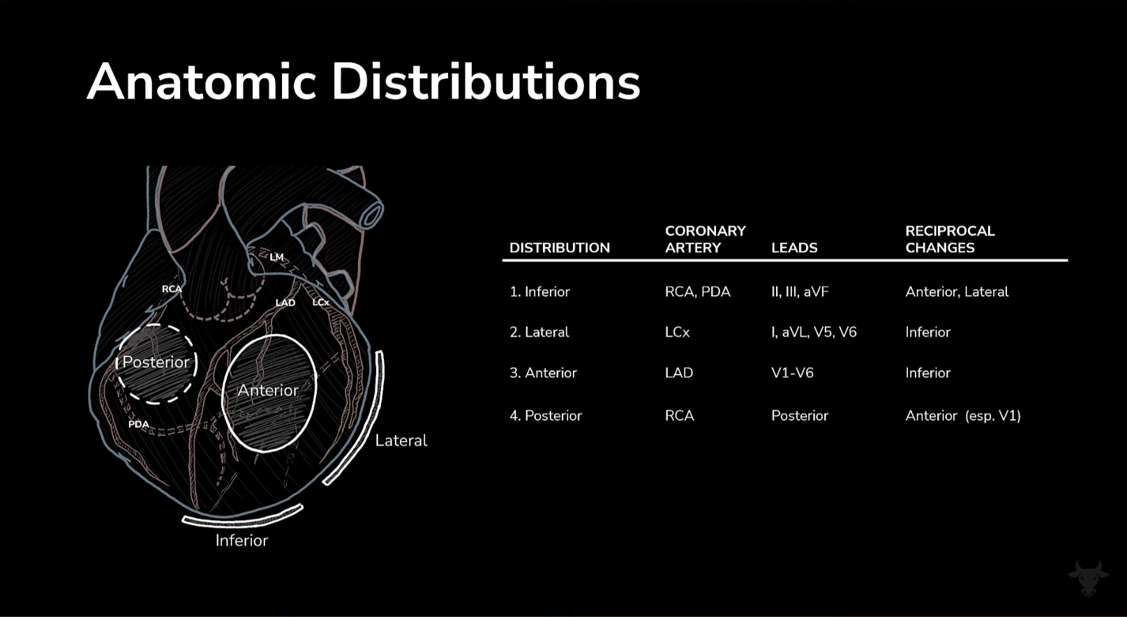
| LEADS | WALL (LV) | ARTERY |
|---|---|---|
| II, III, aVF | Inferior | RCA > LCX |
| I, aVL, V5, V6 | Lateral | LCX |
| I, aVL | High Lateral | LCX branch |
| V1-V2 | Septal | LAD or branch |
| V3-V4 | Anterior | LAD |
| V1-V3 | Posterior (in mirror image – R waves / early transition, ST depression) | RCA |
| V4R – V6R (precordial leads placed on right side) | Right ventricle (usually with II, III, aVF) | RCA |
RCA = right coronary artery; LCX = left circumflex artery; LAD = left anterior descending artery
Hypertrophy Criteria
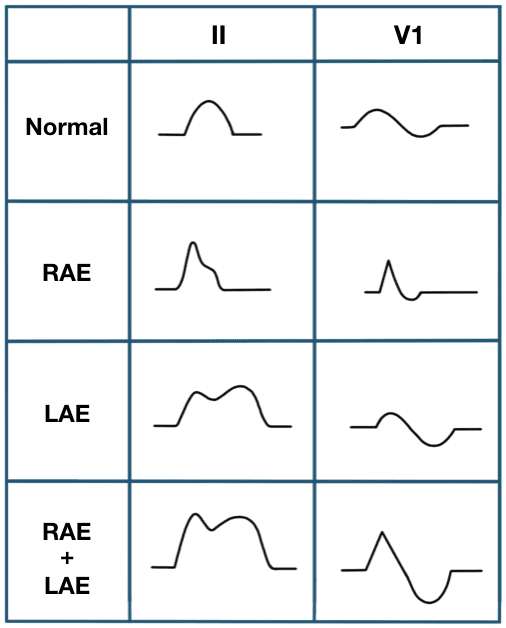
- Atrial – look for biphasic, diphasic, peaked P waves in Leads II and V1
- LAE
- diphasic P wave in Leads II or III, > 120 msec (3 small boxes or more)
- biphasic P wave in V1 with negative portion last that is >40 msec long and >1mm deep (1 small box by 1 small box)
- RAE
- P wave > 2.5 mm tall in Leads II, III, or aVF (2.5 small boxes)
- P wave > 1.5 mm tall in V1 (1.5 small boxes)- Ventricular
- LVH
- Narrow QRS
- S wave in V1 + R wave in V5 or V6 (whichever larger) sums to >35 mm (7 big boxes)
- RVH
- Narrow QRS
- RAD > 110
- R wave in V1 > 7 mm (7 small boxes) or R/S ratio>1
- S wave in V5 or V6 >7 mm or R/S ratio <1
- LVH
- Ventricular
Rhythms and Arrhythmias
AV Blocks
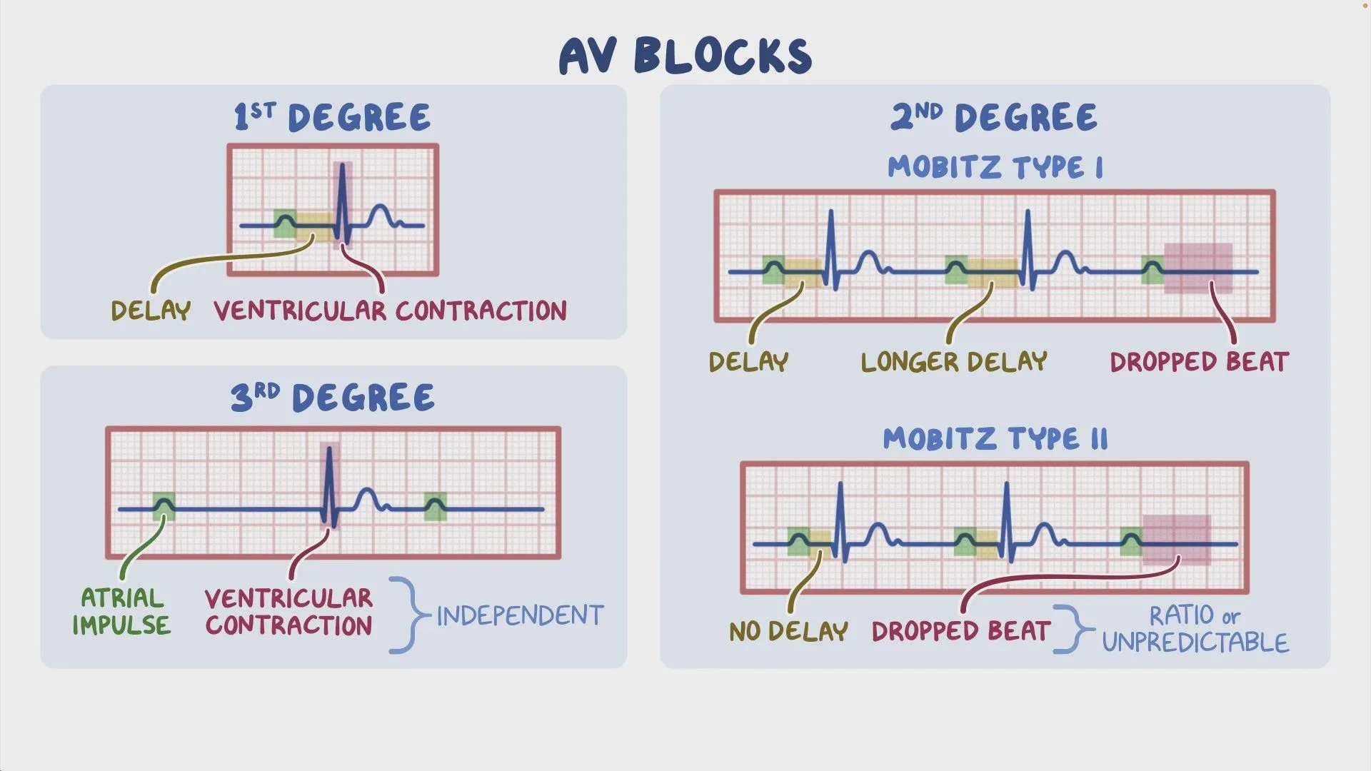
3rd Degree: Need pacemaker!!! Escape rhythm (junctional or ventricular) can cease unexpectedly
Unique Rhythms
| Wellens syndrome | - Deep, symmetric T wave inversions in V2 and V3 OR - Biphasic T waves with terminal negative portion in V2 and V3 |  |
|---|---|---|
| Pulseless electrical activity | - Normal or abnormal heart rhythm while heart not pumping | |
| Brugada syndrome | - RBBB with STE - V1, V2, V3 |  |
| Acute pericarditis | - Classic - STE across most leads - Lack of reciprocal ST changes - PR segment depression in several leads - PR segment elevation in aVR - Late stage - T wave inversion in most leads |  |
| Benign early repolarization | - Widespread upward-sloping STE - Especially V2-V5 - No reciprocal ST depression - Elevated J point with notching or slurring - Prominent, asymmetric T waves that are concordant with QRS |  |
| Pacemakers | - Vertical lines when pacer fires | |
| Single chamber - Atrial pacing | Pacer spike → P wave → QRS |  |
| Single chamber - Ventricular pacing | Pacer spike just before QRS |  |
| Dual chamber pacing |  | |
| Hyperkalemia | - Peaked T waves - QRS eventually widens into a non-perfusing rhythm → looks like sine wave |  |
| Hypokalemia | - Flattened T waves - May see a U wave after T wave |  |
| Hypercalcemia | - Shortens QT interval | |
| Hypocalcemia | - Prolongs QT interval - Watch for torsades |  |
| Hypothermia | - Bradycardia - QT prolongation - Osborne / J waves |  |
| COPD | → possible findings - Multi-focal atrial tachycardia - Atrial fibrillation - Small QRS amplitude - I, aVL, V4-V6 - Right axis deviation - RA and RV hypertrophy |   |
| Pulmonary embolism | → possible findings - Sinus tachycardia - T wave inversion - Inferior and precordial leads - II, III, aVF, V1-V4 - Non-specific ST segment and T wave changes - RAE, RAD, RBBB - Buzzword findings → S1Q3T3 pattern - Lead I → prominent S wave - Lead III → Q wave + T wave inversion |  |
EKG Mechanics
P Wave Basics
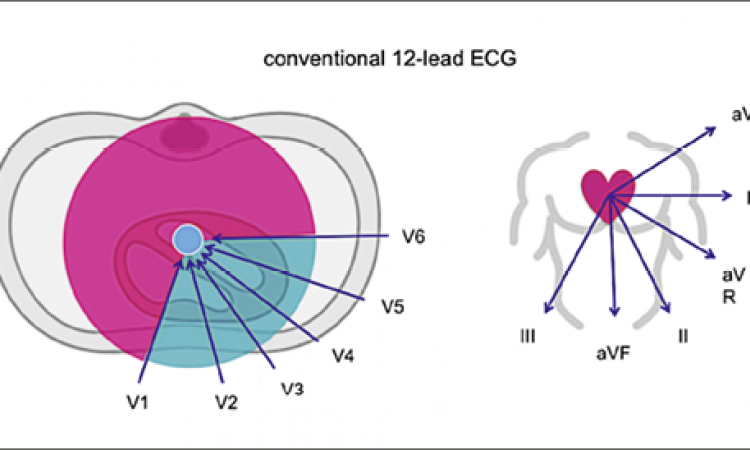
- Peaked P Waves: pulmonary disease (RAE)
- Bifid P Waves: mitral disease (LAE)
- Biphasic P waves: Normal in V1, large terminal component = LAE
Read EKGs the Same Way Every Time
- Go in this order:
- Rate: Count big boxes between QRS complexes (300 ÷ # boxes).
- Normal: 60-100 bpm. •
- Rhythm: Sinus (P before every QRS) vs Irregular (AFib, ectopy)
- Axis: Normal (-30 to +90°)? Check leads I and aVF for QRS direction if it looks like its LAD check lead two to see if its physiologic (thumbs method will do you fine).
- Intervals: PR (120-200 ms), QRS (<120 ms), QTc (<440 ms men, <460 ms women).
- Signs of Ischemia:
- ST changes & Q waves: Check the contiguous leads in order dont just glance at random leads: • Inferior/right heart: II, III, aVF, aVR, V1. • Lateral: I, aVL, V5-V6. • Anterior: V1-V4.
- R-wave progression: Smooth increase across V1-V6. Poor progression suggests prior MI or LVH
- T waves: hyperacute, flattened, inverted
Know STEMI and STEMI Equivalents • STEMI Criteria: ST elevation in 2+ contiguous leads: • ≥1 mm in most leads. • V2-V3: ≥1.5 mm (women), ≥2 mm (men >40), ≥2.5 mm (men <40).
• Remember dynamic MI Changes: Hyperacute T waves → ST elevation → T-wave inversion → Q waves. *Pearl: Pseudo-normalization (T waves normalize in a patient with chest pain) is bad—think evolving MI.
• STEMI Equivalents (memorize these):
• De Winter’s: Upsloping ST depression with peaked T waves in V1-V4 (LAD occlusion).
• Wellens’: Biphasic (Type A) or deeply inverted (Type B) T waves in V2-V3. No stress test—needs cath!
• Left Main: Diffuse ST depression with ST elevation in aVR.
• Sgarbossa: STEMI in LBBB (Google the criteria but you better know it baby!)
• Posterior MI: ST depression in V1-V3 (get posterior leads).
• Right-Sided MI: ST elevation in V4R (use right-sided leads).
For patients with Syncope/Dizziness run through these patterns after your systematic read from above: • HOCM: LVH, deep Q waves, dagger-like Qs in lateral leads.
• Brugada: RBBB-like pattern with ST elevation in V1-V2.
• WPW: Short PR, delta waves.
• Long QT
• High-Grade Blocks: 2nd-degree type II or 3rd-degree block.
• ARVC: Epsilon waves, T-wave inversion in V1-V3.
• Ectopy: Frequent PVCs or VT.
Electrolyte Changes you should know:
• Hyperkalemia: Bradycardia, Peaked T waves → widened QRS → dropped P waves → sine wave. *Pearl: If it looks like “slow VT” remember thats not a thing lol. Ventricular tachycardia is fast not slow silly goose! —treat with calcium gluconate bc if you give amio its a clean kill take an ear and add another notch killer
• Hypokalemia: U waves, flat T waves, ST depression, prolonged QT.
• Hypercalcemia: Short QT.
• Hypocalcemia: Long QT.
• Hypomagnesemia: Long QT, flat T waves, widened QRS (severe).
• Hypermagnesemia: Long PR, mimics hyperkalemia.
Tox and Miscellaneous Pearls • Digoxin: Salvador Dalí pattern (scooped ST segments, this is just dig use not toxicity!!), slow Afib think dig , or bidirectional VT thats classic dig toxicity but rare. Atrial tachycardia with AV block classic dig toxicity.
• TCA Overdose: Wide QRS, large terminal R wave in aVR.
• Pericarditis: Diffuse PR depression, widespread ST elevation. (Pearl* if its myocarditis obs your admitting but nsaids are contraindicated)
• Tamponade: Tachycardia, low voltage, electrical alternans.
• PE/Right Heart Strain: S1Q3T3, T-wave inversion in inferior (II, III, aVF) or V1-V2, ST elevation in aVR. T-wave inversion in inferior + right precordial leads is most specific ecg sign for right heart strain btw (thats on boards)
• Aneurysm or even Takotsubo: Persistent ST elevation with deep Q waves post-MI.
• Benign Early Repolarization: Concave ST elevation, J-point slurring, upright slightly asymmetric T waves (benign in healthy patients and most notable in v4-v6)
• Pathologic Q Waves (know what defines them ie: >40 ms wide or >25% of R-wave height. Buuuuuut Q waves in V1-V3 are always pathologic even if they arnt wide or deep.
• Inverted U Waves: Specific for myocardial ischemia if you see these in a pt w chest pain he stays for stress testing. Honestly you should get in the habit of checking for this when assessing the dynamic changes for MI routinely
• Slow AFib: Think hypothyroidism, digoxin, or hyperkalemia.