Potassium
Hypokalemia:
Much more well tolerated than hyperkalemia
- Main causes: GI losses, Renal Losses, Intracellular shifts, Low K Intake
- GI Losses: Diarrhea, vomiting, NG tube, etc.
- Renal losses: diuretics, nephrotic syndrome, etc
- Intracellular shifts: medications (insulin, beta agonists, etc)
- Low K intake: rare, alcoholics/anorexia
Potassium Repletion
- LOOK AT PHOSPHORUS
- 3<K<3.5 and asymptomatic: oral (if able)
- <3.0: IV potassium
- K goes up .1 every 10 meq
- Max repletion rate: 1 meq /kg / day
- How to order: e.g. 10 meq in 50mL ran over 1 hour (times how much K needs to increase)
- PIV: 10meq/hr
- Central line: 20meq / hr
- IV potassium burns veins
Hyperkalemia:
- Main causes: hemolysis of lab sample, acidosis, medications, decreased renal excretion, excessive intake
- Hemolysis: always ask “does this make sense”
- Acidosis: pH<7.35 causes shift extracellular
- Meds/Decreased renal excretion: ACE-I, MRA, Beta Blockers, AKI/CKD
- Excessive intake: ate too much! Rare
Total Bilirubin / AST / ALT / ALP / GGT :
Important functional measures of hepatobiliary system
Patterns:
R Factor for Liver Injury = points towards cholestatic vs. hepatocellular vs mixed injury
Cholestatic
- increased ALP and GGT
Mixed: - increased AST/ALT and ALP
- think DILI, late viral hepatitis, early biliary obstruction
Hepatocellular: - viral/autoimmune hepatitis, hemochromatosis, Wilson’s disease, alpha 1 antitrypsin deficiency
- AST predominant: alcohol / cirrhosis
Bilirubin
Elevated bilirubin should be fractionated to indirect (unconjugated) and direct (conjugated)
Abnormal concentrations may indicate cholestasis or hepatocellular damage in the presence of other abnormal liver tests
- Jaundice = Tbili > 2.5-3.5
- Direct = conjugated
- Conjugated bili = pruritis
Vocab: - kernicterus (bilirubin encephalopathy) - brain damage causes by newborn bilirubin buildup
- unconjugated bili > 25
AST / ALT
- markers of hepatocellular injury
- ALT more specific to liver
- Generally, hepatocellular injury results in an AST:ALT ratio >1. However, alcoholic liver disease usually presents with a ratio of ≥2. This ratio may also be elevated in cases of NAFLD, cirrhosis, or Wilson disease, but the elevation is usually not >2
Clinically significant elevations:
The American College of Gastroenterology defines the following categories for AST and ALT elevations:
• Borderline: <2× ULN
• Mild: 2–5× ULN
• Moderate: 5–15× ULN
• Severe: >15× ULN
• Massive: >10,000 IU/L
Borderline and mild elevations (<5× ULN) are most commonly seen in metabolic dysfunction-associated steatotic liver disease (MASLD, formerly NAFLD), alcoholic liver disease, chronic viral hepatitis, and drug-induced liver injury.
Moderate elevations (5–15× ULN) may be seen in acute viral hepatitis, drug-induced liver injury, or autoimmune hepatitis.
Severe and massive elevations (>15× ULN, especially >10,000 IU/L) are typically associated with ischemic hepatitis (shock liver), severe drug-induced/toxic hepatitis (e.g., acetaminophen toxicity), or acute viral hepatitis. Non-hepatic causes such as rhabdomyolysis can also result in very high AST levels.
Alk Phos / GGT
Elevation of ALP is suggestive of cholestasis
Serum 1/2 life: 7 days
- can take 1 week to decrease following resolution of biliary obstruction
GGT and/or 5’NT may be useful to identify whether ALP elevation has a hepatic origin
ALP also found in:- Liver
- Bone
- Kidney
- Intestine
- Neutrophils
GGT:
- GGT measurement can be used to confirm the origin of elevated ALP
- Elevated GGT indicates a hepatic cause of ALP elevation
- GGT is often used to evaluate alcohol use, but it is nonspecific and may be elevated with diseases such as NAFLD and other hepatobiliary disorders
Causes of elevated ALP
- Biliary obstruction (intra/extra hepatic)
- Other liver pathologies: cancer, DILI, infections, etc
- Cancer
- Pregnancy (fetal isoenzyme)
- AIDS
- Other: intra-abdominal infections, cholestasis of sepsis, Hodgkin lymphoma, myeloid metaplasia, and osteomyelitis
- Leukemoid reaction
Synthetic Function
Albumin
- Liver disease decreases albumin synthesis and decreases serum albumin concentration
- In the absence of other markers of liver disease, low albumin concentration may be related to malnutrition or protein loss (e.g., malabsorption)
PT:
- Elevated PT that is unresponsive to vitamin K supplementation is suggestive of poor liver function
Calcium:
general
total calcium is a screening test only - ionized calcium is what matters physiologically
- 8.5-10.5 mg/dL (or 2.12-2.62 mM) is the normal range.
- Total calcium is not an accurate predictor of ionized calcium (since ionized calcium depends on numerous factors including pH, albumin level, sodium level, and phosphate level).
- iCal measures the biologically active calcium. This is the most accurate and preferred way to measure calcium.
- 1.1-1.3 mM is the normal range seen in healthy people, but most critically ill patients tend to run below this range.
- <0.8 mM probably warrants treatment (although there is no clear evidence that treatment improves outcomes).
- <0.65 mM is critically low (possibly causing hypotension)
Serum calcium = ionized calcium + bound calcium (albumin)
Interpret # in context of albumin
- basically impossible to be hypocalcemic if you have bones and a working PTH axis
- HOWEVER:
- Ca is albumin bound
- low albumin = low total calcium (but not functional aka ionized Ca)
ST segment duration is inversely related to the ionized calcium level
- Hypercalcemia causes ST segment shortening
- Hypocalcemia causes ST segment prolongation.
- This may be noticed as QT prolongation with a normal-sized T-wave.
- This may be seen on the patient’s monitor or on a rhythm strip (without a full EKG).
- Hypocalcemia may cause torsade de pointes.
Hypocalcemia:
Causes:
- hypoparathyroidism
- impaired vitamin d synthesis / action
- inadequate intake
- malabsorption
- liver disease / CKD
- hypomagnesemia
- calcium chelation / precipitation
- Hyperphosphatemia of any etiology, for example:
- Renal failure.
- Tumor lysis syndrome.
- Rhabdomyolysis.
- Exogenous citrate:
- Massive transfusion (especially if combined with hepatic/renal dysfunction).
- Plasmapheresis and leukapheresis (fresh frozen plasma contains citrate).
- Renal replacement therapy with citrate regional anticoagulation.
- Pancreatitis (may relate to systemic inflammation and release of free fatty acids).
- Poisoning:
- Ethylene glycol poisoning
- Hydrofluoric acid.
Chvostok’s Sign: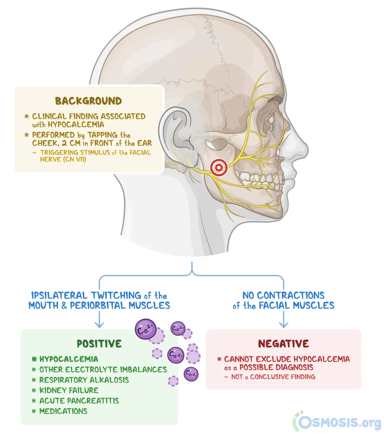
Trousseau Sign: inflate a blood pressure cuff to 20 mm Hg above the systolic blood pressure for 3 minutes to elicit carpal spasms because of the increased excitability caused by local ulnar and median nerve ischemia.
Albumin:
Important synthetic measure of liver - is the liver producing the things it’s supposed to?
(Note- clotting factors/fibrinogen are often preserved or even increased in low albumin states)
Causes of low albumin:
- #1: infection/inflammation (negative acute phase reactant)
- CKD / nephrotic syndrome (>3.5g / day)
- Damaged liver (cirrhosis, hepatitis, surgical resection, etc)
- Malnutrition
Sodium:
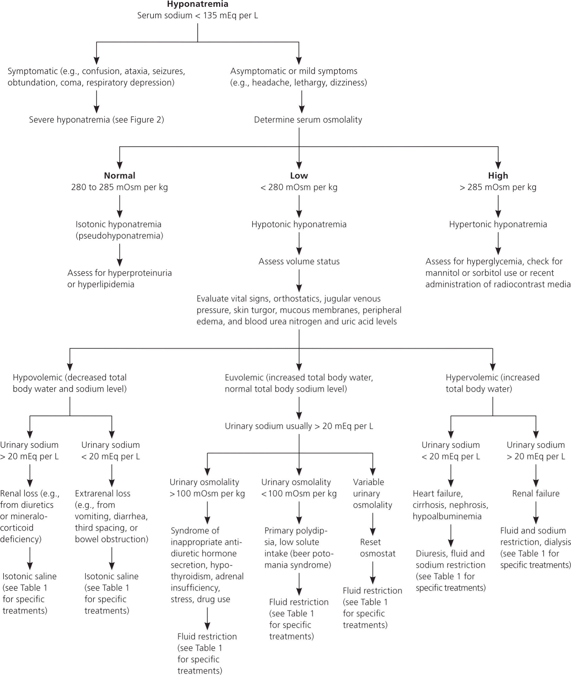
Hyponatremia
- MC is hypotonic (90%)
- MC Cause inpatient: fluids (iatrogenic hypervolemic hyponatremia)
- Urine Studies:
- uOsm: >100= ADH is working, <100= ADH is not working
- uNa: <20=kidneys are working, >20=kidneys are losing Na
- Urine Studies:
- Hypotonic Hyponatremia: 90% of cases
- Hypovolemic:
- GI losses, 3rd spacing, Diuretic meds
- uNa<20: GI losses, 3rd spacing
- uNa>20: Diuretic meds
- GI losses, 3rd spacing, Diuretic meds
- Euvolemic:
- SIADH
- uOsm>100: SIADH, hypothyroidism, Adrenal insufficiency
- uOsm<100: “tea and toast” diet, beer potomania, primary polydipsia
- SIADH
- Hypervolemic:
- Treatments:
- Hypovolemic: Fluids (NS)
- Euvolemic: Fluid Restriction
- Hypervolemic: Diuresis
- Hypovolemic:
Hypernatremia
- Free Water Deficit
- AI/DI
Hemoglobin:
- RBC life = 120 days
- Rapid changes = bleeding or hemoconcentration/dilution
- Do you suspect bleeding?
- Look for signs in foley, drain, poop, vomit, POCUS, etc
- Cross reference with fluid administration and urine/drain output
- Changes in Hgb concordant with volume status = likely not bleeding
- Do you suspect bleeding?
- Urine Output
- Weight (kg) x 0.5 = mL/hr
- Average adult: 30mL/hr or 720mL/day
- 1L/day = 42mL/hr = 84kg patient (185lbs)
Creatinine:
- complete anuria will raise Cr by 1.0 mg/dl per day
- not elevated until ~%50 reduction in renal function
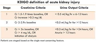
- KDIGO
BUN
- End product of protein metabolism
- 85% eliminated by kidneys, 15% by GI tract
- Increases if renal elimination impaired (AKI, CKD), GI bleeding, high protein diet, severe liver disease
- Increases earlier in chronic renal dysfunction than SCr
Ammonia
Hepatic Encephalopathy
- not indiciative of degree of encephalopathy