quick reference
Scores:
HEART Score for Major Cardiac Events
GRACE ACS Risk and Mortality Calculator
IBCC
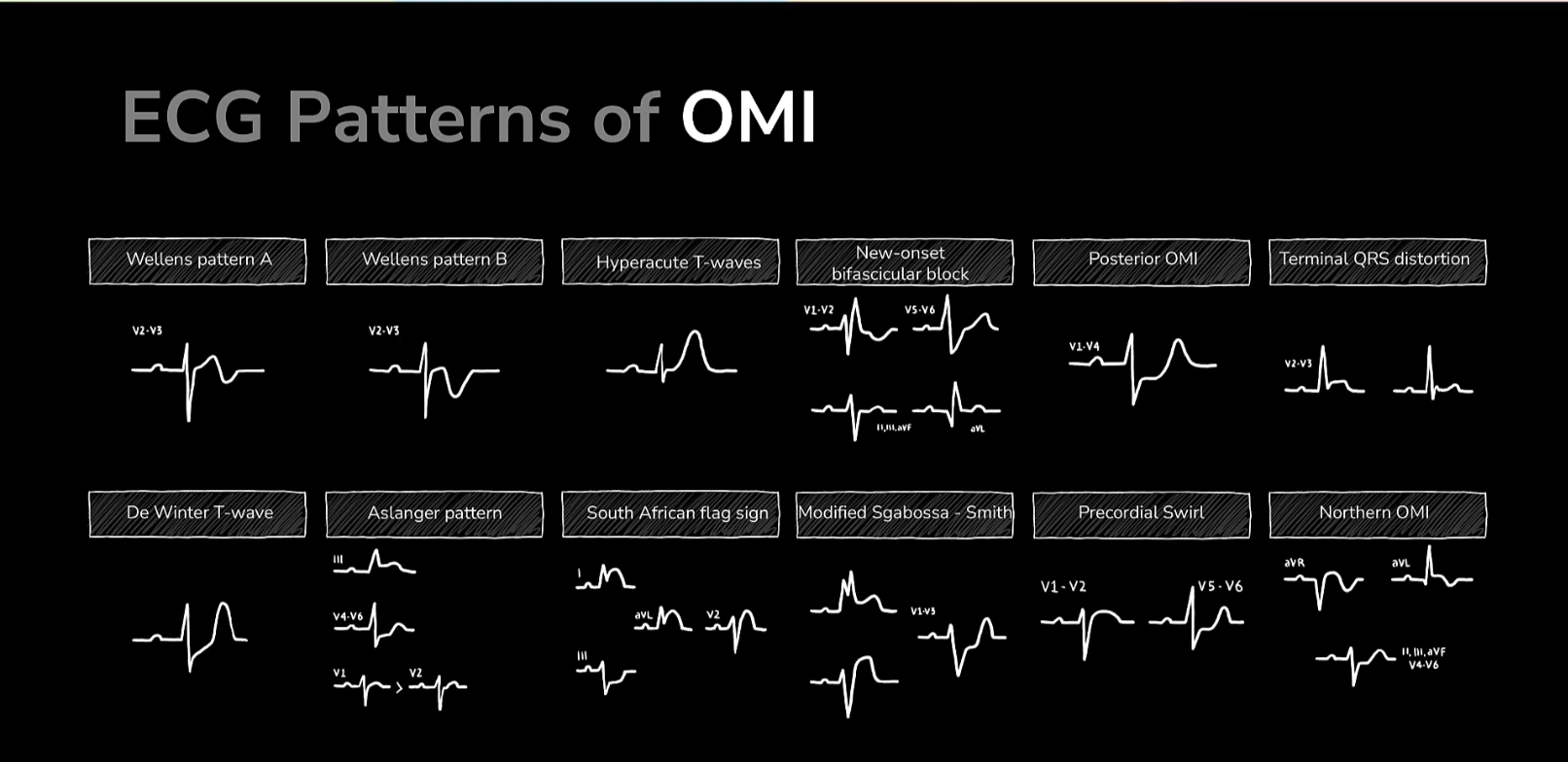
Type-1 MI (OMI & NOMI) and related complications
2025 AHA Guidelines:
2025 Guideline for the Management of Patients With Acute Coronary Syndromes
clinical features
chest discomfort—typically described as:
- pressure, heaviness, tightness, or pain, often substernal and possibly radiating to the arms, neck, jaw, or back.
- This discomfort is usually of sudden onset, may occur at rest or with minimal exertion, and lasts more than a few minutes.
- Diaphoresis!
also look for other common features:
-
dyspnea, nausea, vomiting, palpitations, syncope, dizziness, unexplained fatigue, or epigastric discomfort
-
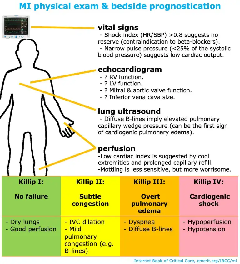
Physical findings may include signs of heart failure (e.g., pulmonary rales, S3 gallop), hypotension, tachycardia, or arrhythmias
-
Risk factors include:
- Personal history of atherosclerotic disease (e.g., prior MI, peripheral artery disease).
- Age.
- Smoking.
- Diabetes, hypertension.
- Radiation therapy.
- Sympathomimetic use (cocaine/methamphetamine).
- Vasculitis.
-
Likelihood ratio for pain attributes: (evidence)
- Radiation to arm(s) or shoulder(s): LR ~4.
- Worse with exertion: LR ~2.5
- Associated with diaphoresis: LR ~2.
- Sharp: LR ~0.3
- Positional: LR ~0.3
- Reproducible with palpation: LR ~0.3
- Pleuritic: LR ~0.2
- Caution: chest pain may be atypical in women, elderly, or patients with diabetes.
workup
Initial:
- EKG, vitals, telemetry, 2x IV access
Diagnostics:
- CBC
- BMP
- CBG
- Chest X-ray
- Troponin
- PT-INR + PTT, Type & Screen
- +/- lactate
- lipid panel
diagnosis
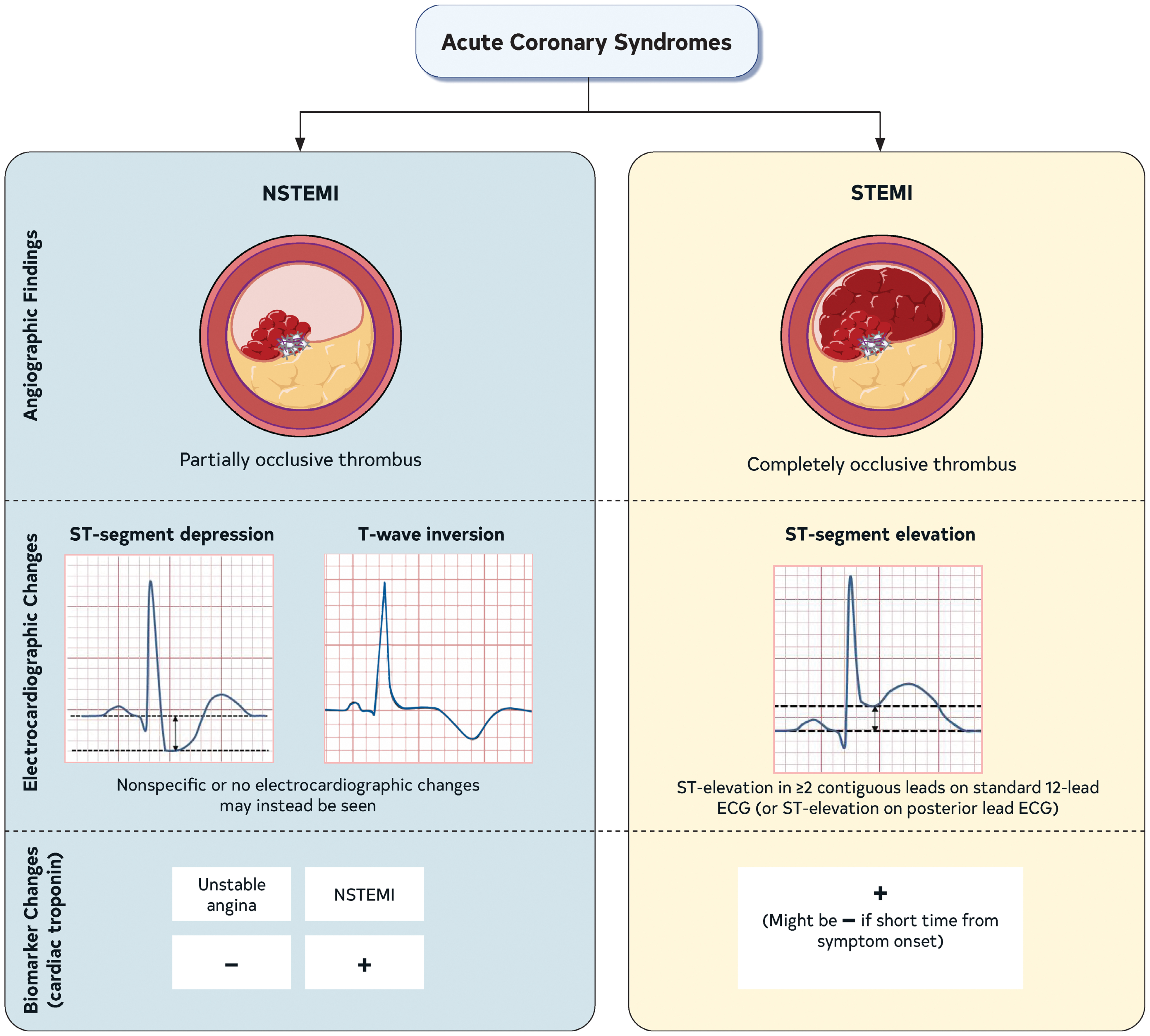
EKG + hsTrop
| NSTE-ACS | STEMI | |
|---|---|---|
| Electrocardiographic evidence of ischemia | New or presumed new and usually dynamic horizontal or down-sloping ST-segment depression ≥0.5 mm in ≥2 contiguous leads and/or T-wave inversion >1 mm in ≥2 contiguous leads with prominent R wave or R/S ratio >1 or transient ST-segment elevation. | New or presumed new ST-elevation of ≥1 mm in ≥2 anatomically contiguous leads (measured at the J-point) in all leads other than V2-V3 and ≥2 mm in men ≥40 y, ≥2.5 mm in men <40 y, and ≥1.5 mm in women regardless of age in leads V2-V3.* |
| Other observed electrocardiographic changes | Many patients with NSTE-ACS have either nonspecific ST-segment or T-wave changes or a normal ECG. The absence of electrocardiographic evidence of ischemia does not exclude ACS. | Posterior leads (V7-V9) should be obtained in patients with suspected left circumflex occlusion particularly in the setting of isolated ST-segment depression ≥0.5 mm in leads V1-V3. |
treatment
medical management
Initial:
- Morphine: Avoid if possible, inhibits P2Y12 activity
- O2: if SpO2 <90%
- Nitroglycerin .3mg every 5 min (max of 3)
- no PDE-4 (viagra, etc) w/in 24hrs, tadalfil w/in 48hrs,, caution if SBP <110, contraindicated in RV involvement (inferior MI)
- Aspirin: 324mg chewed
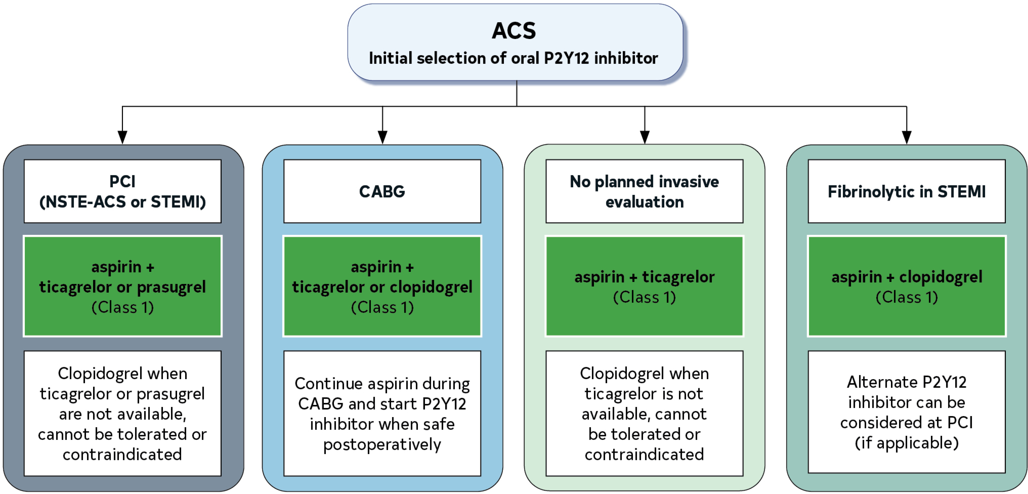
- P2Y12: yes, post-PCI (may delay CABG surgery)
- Heparin Bolus + Drip:
- Initial therapy: Loading dose 60 IU/kg (max 4000 IU), with initial infusion 12 IU/kg per h (max 1000 IU/h) adjusted to therapeutic aPTT range of 60-80 s.
- To support PCI: In patients who have received prior anticoagulant therapy, additional UFH as needed to achieve an ACT 250-300 s.
- In patients who have not received prior anticoagulant therapy, 70-100 U/kg initial bolus to achieve target ACT of 250-300 s.
- With fibrinolytic therapy: Loading dose 60 IU/kg (maximum 4000 IU) with initial infusion 12 IU/kg per h (maximum 1000 IU/h) adjusted to therapeutic aPTT range.*
Within 24hrs:
- Beta Blocker
- ACE-i/ARB (if LVEF<40%, HTN, T2DM, anterior STEMI)
- High Intensity Statin
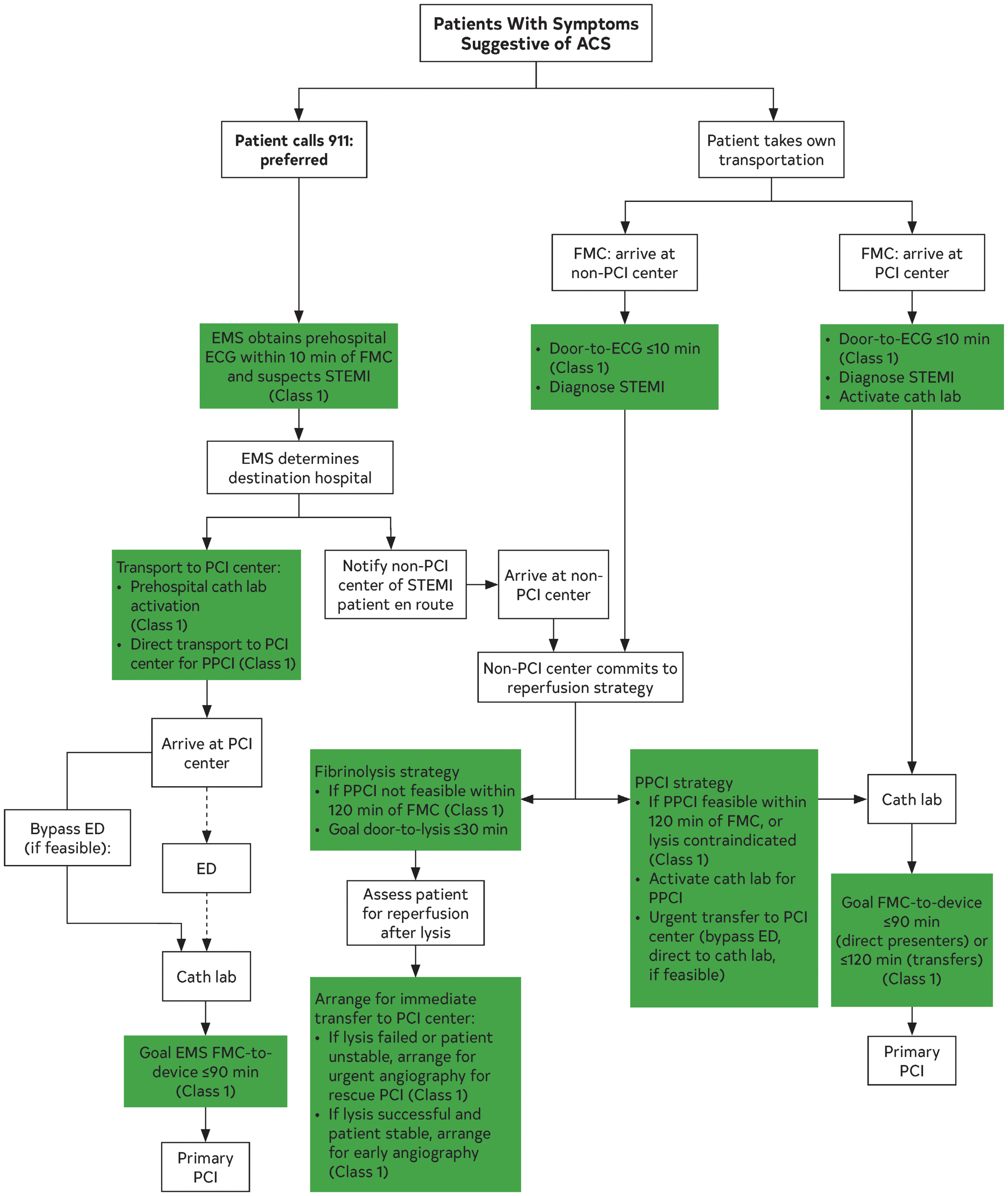
reperfusion
PCI
PCI-capable Center: <90min door-to-balloon (correct terminology: FMC to device activation)
If able to transfer in time: <120min door-to-balloon
fibrinolytics
If unlikely to achieve PCI within 120 min:
- Patients should be transferred to a PCI-capable hospital after initiation of fibrinolytic therapy for routine coronary angiography
| Fibrinolytic Agent | Dose |
|---|---|
| Tenecteplase (TNK-tPA) | Single IV weight-based bolus* |
| Reteplase (rPA) | Two 10-unit IV boluses given 30 min apart (administered over 2 min) |
| Alteplase (tPA) | 90-min weight-based infusion† |
| Consent |
- Most important part of decision to use fibrinolytics
| Fibrinolytic Contraindications |
|---|
| Absolute Contraindications |
| Any prior ICH |
| Known structural cerebral vascular lesion (eg, arteriovenous malformation) |
| Known malignant intracranial neoplasm (primary or metastatic) |
| Ischemic stroke within 3 mo except acute ischemic stroke† |
| Suspected aortic dissection |
| Active bleeding or bleeding diathesis (excluding menses) |
| Significant closed-head or facial trauma within 3 mo |
| Intracranial or intraspinal surgery within 2 mo |
| Severe uncontrolled hypertension (unresponsive to therapy) (SBP >180 mm Hg or DBP >110 mm Hg) |
| Relative Contraindications |
| History of chronic, severe, poorly controlled hypertension |
| Significant hypertension on presentation (SBP >180 mm Hg or DBP >110 mm Hg) |
| History of prior ischemic stroke >3 mo |
| Dementia |
| Known intracranial pathology not covered in absolute contraindications |
| Traumatic or prolonged (>10 min) CPR |
| Major surgery (<3 wk) |
| Recent (within 2 to 4 wk) internal bleeding |
| Noncompressible vascular punctures |
| Pregnancy |
| Active peptic ulcer |
| Oral anticoagulant therapy |
reperfusion algorithm